
Cracked Tooth Imaging: Why We Capture Before, During and After Treatment
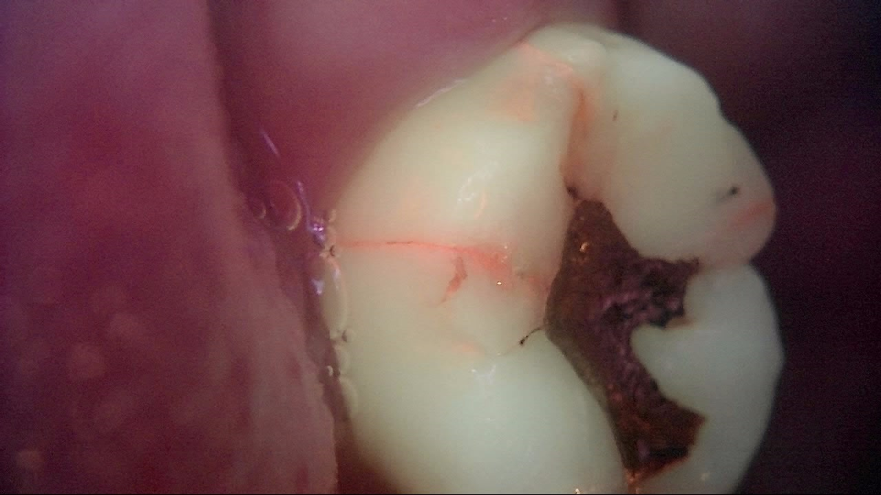
"It hurts when I bite — but I can't tell you which tooth." That is how most cracked tooth visits begin. The bite test is equivocal, the radiograph is clean, and from the visual exam alone you can't tell a harmless craze line from a crack that runs into dentin. Cracked tooth imaging with Qray fluorescence (405 nm) won't make the call for you — it gives you something to look at, record and show the patient. Here is how to fold crack visualization into the appointment, before, during and after treatment.
Why is a cracked tooth so hard to confirm?
Cracked tooth symptoms are sporadic and hard to reproduce, and the crack itself rarely shows on radiographs or under normal light. A Gangnam Severance Hospital team (Lee et al., Photodiagnosis Photodyn Ther 2023) makes exactly this point, noting that clinicians have relied on the operating microscope, transillumination and dyes such as methylene blue to make crack lines visible.
The AAE classification is still the frame of reference. It sorts longitudinal fractures into five types, each with a different origin, symptom profile and prognosis. Knowing which type you are dealing with — and how far it extends — drives the treatment plan.
| AAE type | Origin | Typical symptoms | Prognosis |
|---|---|---|---|
| Craze line | Crown | None | Excellent |
| Fractured cusp | Crown | Mild, mainly on biting and cold | Good |
| Cracked tooth | Crown ± root | Acute pain on biting, sometimes sharp pain to cold | Questionable — depends on depth and extent |
| Split tooth | Crown + root | Marked pain on chewing | Poor unless the crack ends just subgingivally |
| Vertical root fracture | Root | Vague pain, mimics periodontal disease | Poor |
One proposed addition: our case file adds a "craze line with red fluorescence" row with a questionable prognosis, even without symptoms — a suggestion from clinical observation, not an accepted classification.
What does a crack look like under Qray fluorescence?
A crack line reads as a bright line; once biofilm enters the gap, it turns red. The green fluorescence coming up from dentin tends to escape more brightly through the gap of a crack, so an early craze line often appears as a light line. When biofilm moves into that space, red fluorescence appears. The same crack line, if it shows red, suggests that more space has opened.
The red is a bacterial signal. Lee et al. cite histology showing bacterial colonization along crack lines and the surrounding dentinal tubules, and explain that the red fluorescence of porphyrins left by those bacteria helps locate the crack. For the underlying physics, see Quantitative Light-Induced Fluorescence: What 405 nm Shows.
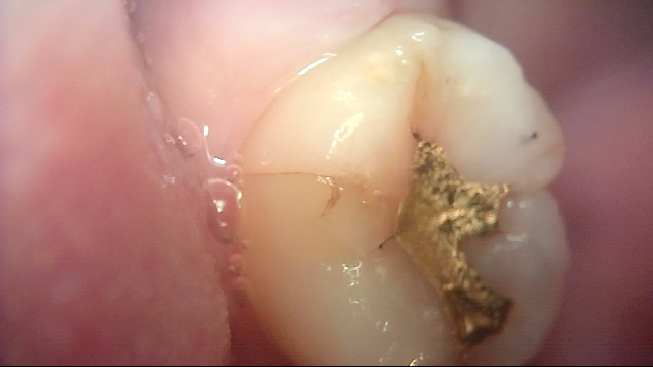
Under white light, you see a faint hairline. Under Qray fluorescence, the same line reads as a continuous red trace across the cusp.
What does the research show about crack visualization?
In a clinical study, 84 of 90 cracked teeth showed their crack lines as red fluorescent lines. Lee et al. (2023) used Qraypen C to image 90 teeth from 66 patients clinically diagnosed with cracked teeth, including asymptomatic contralateral teeth with visible crack lines. In 84 teeth, the crack line showed as a red fluorescent line above the analysis software's cut-off. In some teeth, fluorescence revealed additional crack lines that had not been seen on visual inspection. Teeth that were tender to percussion showed significantly greater fluorescence loss and red fluorescence along the crack line. The authors concluded that QLF is a useful assistive tool for cracked teeth — an adjunct, not a replacement.
An extracted-tooth study found that red fluorescence differed by fracture type. Ku et al. (Photodiagnosis Photodyn Ther 2022) examined 33 extracted teeth and found red fluorescence in 82% of cracked teeth and 83% of split teeth, but in none of the vertical root fractures. Because these were extracted teeth, treat this as evidence of mechanism, not chairside performance.
Read the limits, too. Lee et al. note that chairside distance and angle vary, and light angle affects the analysis. Fluorescence does not replace the bite test, percussion, cold testing or radiographs — it adds visible evidence to them.
Why capture before, during and after treatment?
Some cracks only show once the restoration is out or the access is open. Son et al. (J Endod 2021) used Qraypen C before and after restoration removal and inside the access cavity, and reported that the images helped locate cracks and gauge their depth during treatment. In a 2023 case report by the same group, removing a ceramic restoration with no visible findings revealed a distinct red crack line that continued into the pulp chamber; the team removed as much of it as possible, guided by the fluorescence images.
In one clinical case, a lower second molar followed the same arc. The patient reported discomfort when eating. The Qray fluorescence image showed red on #18 (#37 in FDI), and the radiograph suggested shallow dentin caries. A closer capture revealed a vertical crack line along the DEJ. During crown prep, removing the red areas uncovered dentin caries far deeper than the radiograph suggested. Because the crack was heading from crown toward root, the tooth was restored with a crown and is being monitored.
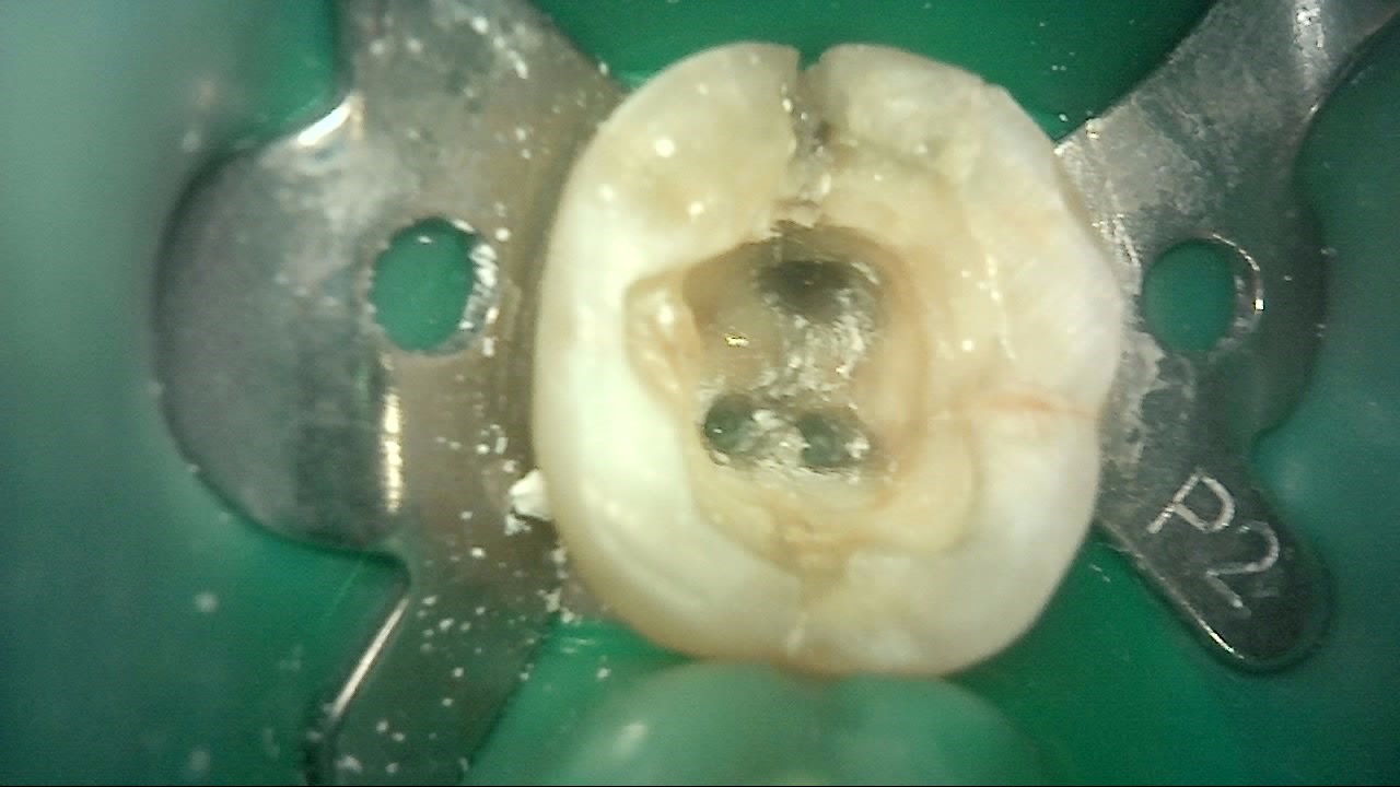
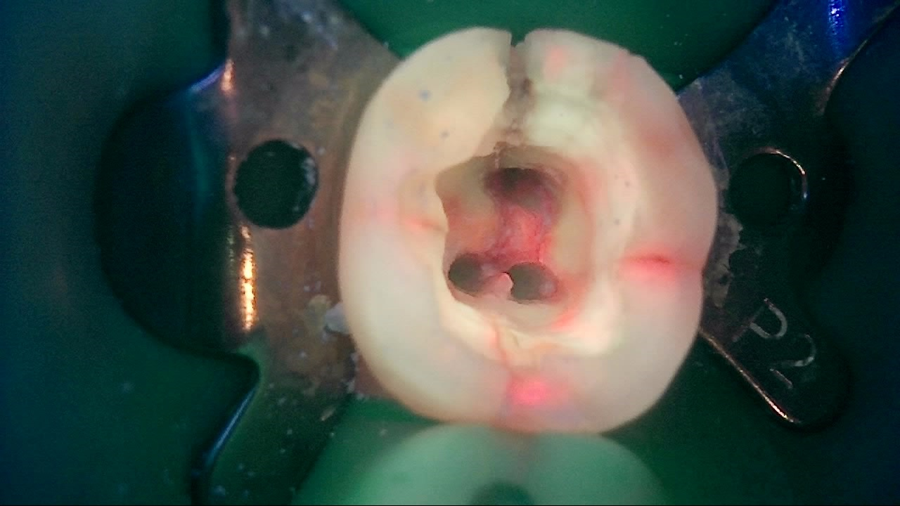
With the access open, a second capture shows crack lines running across the cavity walls and marginal ridge in red.
Capture tips that experienced users often share:
- Capture at every stage — before treatment, during prep and at the margin after reduction — to see how far the line runs.
- When you suspect a crack, capture both occlusal and palatal/lingual views, and angle the pen to follow the line toward the root.
- Brace Qraypen C on the opposing tooth for sharper focus.
- Mark occlusal contacts with articulating paper first, then capture Qray fluorescence. Contacts and crack lines often line up, which makes the conversation with the patient much simpler.
Where does imaging fit in a cracked tooth workup?
Qray fluorescence imaging is the documentation step between reproducing symptoms and treating. A cracked tooth workup is commonly organized into five steps.
| Step | What you do | Where imaging fits |
|---|---|---|
| 1. Listen to the patient | Character, timing and location of pain | — |
| 2. Reproduce the symptoms | Percussion, bite, cold, EPT | Narrow to the suspect tooth, then capture |
| 3. Document the crack | Staining, periodontal probing | White-light + Qray fluorescence pair to record location and extent |
| 4. Treat by the condition of the tooth | Restoration, RCT, crown | Recapture during treatment to check extension |
| 5. Regular check-ups | Follow at 3 and 10 years | Recapture under the same settings and compare |
A similar four-step approach is also used in practice: find the problem tooth with quantitative light-induced fluorescence, reproduce the chief complaint by having the patient bite on a cotton roll in several directions, assess how much bacteria has entered the crack, then judge prognosis from probing depth, pulp vitality and crack location. Explaining the prognosis up front helps avoid misunderstandings later.
What's next
A crack image keeps its value after the appointment. Record it per tooth in LINKDENS, tag it, and at the next recall you can recapture the same tooth under the same settings and see whether the line has changed. Sent as a report, it lets the patient see at home what "a crack in your tooth" means. When patients see the crack line, they tend to accept occlusal adjustment more readily, and it can give the team a natural moment to discuss night guards and other care before the tooth needs endo or a crown. For a practice owner, a cracked tooth becomes something you track, not a one-time fix — and carried forward through QBLISScare, treatment becomes care.
FAQ
Q. Can a Qray device diagnose a cracked tooth?
A. No. Qray devices make crack lines and the biofilm inside them visible, which helps you confirm and document what you suspect. The diagnosis stays with the dentist, together with symptom testing and radiographs.
Q. Does every red-fluorescing crack need treatment?
A. No. Red means biofilm has settled into the gap. Read it alongside symptoms, probing depth and pulp status, then decide whether to monitor, manage occlusion or restore.
Q. Which device should I use for cracked tooth imaging?
A. Many teams start with Qraycam Pro for the full-arch overview and use Qraypen C for the suspect tooth and for recapture during treatment. See Qray Intraoral Camera Lineup: Which Device, When?
References
- Lee JI, Jeon MJ, de Josselin de Jong E, et al. Evaluation of the clinical efficacy of quantitative light-induced fluorescence technology in diagnosing cracked teeth. Photodiagnosis Photodyn Ther. 2023;41:103299. DOI
- Ku HM, Kim E, Kim BI. Red fluorescence for assessing longitudinal tooth fractures. Photodiagnosis Photodyn Ther. 2022;38:102845. DOI
- Son SA, Kim JH, Park JK. The effectiveness of a quantitative light-induced fluorescent device for the diagnosis of a cracked tooth: a case report. J Endod. 2021;47(11):1796-1800. DOI
- Son SA, Kim JH, Park JK. Clinical applications of a quantitative light-induced fluorescent (QLF) device in the detection and management of cracked teeth: a case report. Photodiagnosis Photodyn Ther. 2023;43:103735. DOI