
Quantitative Light-Induced Fluorescence: What 405 nm Shows
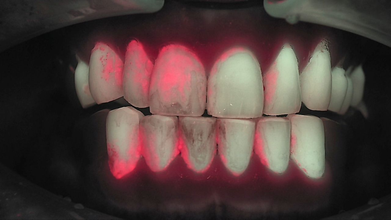
What does quantitative light-induced fluorescence actually show you? If you have ever used a disclosing agent to show a patient their plaque, you know the trade-off: the patient sees the stain, but also leaves with purple lips, and the result is gone after the next rinse. QLF answers the same question with light alone, and it keeps the answer as an image you can compare at the next visit. This post is the anchor for our QLF topic hub. It covers how the signal is generated, what "quantitative" means, and how QLF fits alongside radiographs in a US practice.
What does QLF see under 405 nm light?
QLF relies on one simple fact: tooth and bacteria glow in different colors under 405 nm light. Dental hard tissue is relatively stable and structurally simple. It absorbs high-energy 405 nm excitation light and re-emits part of that energy as green fluorescence. A proprietary optical filter renders the image in colors close to the tooth's natural appearance, so patients recognize their own teeth.
Pathogenic biofilm responds differently. Many late-colonizing bacteria carry porphyrins — complex molecular structures that redistribute much of the absorbed energy internally and emit the rest as red fluorescence. Put simply, red fluorescence appears where porphyrins are present — a natural metabolic byproduct of these bacteria.
| What the 405 nm light hits | What you see | What it means |
|---|---|---|
| Sound enamel and dentin | Green fluorescence (natural tooth color after filtering) | The tooth's own fluorescence |
| Early demineralization | Darker area — fluorescence is lost | Mineral loss, recorded as the degree of fluorescence loss |
| Biofilm matured 3+ days | Red fluorescence | Bacterial porphyrins, recorded as the degree of red fluorescence |
| Crack line | Bright line; red where biofilm has entered the gap | Red along a crack suggests an open space colonized by bacteria |
Once your team knows what each color means, a fluorescence image stops being a pretty picture and becomes data you can read chairside.
Why does plaque fluoresce red — and why "three days"?
Red fluorescence is not an alarm. It is an objective signal that biofilm has been sitting there long enough to mature. Biofilm forms in stages. Early colonizers attach first; later colonizers move in as the community matures and the balance shifts. The later colonizers are the ones that carry porphyrins — the same family of structures as hemoglobin in blood and chlorophyll in plants. Older biofilm is rich in protoporphyrin IX.
Balanced, healthy biofilm does not glow red. Red fluorescence is generally not seen in the symbiotic early-colonizer stage; it tends to appear after the community tips into dysbiosis. That is why red is useful: it points to where the balance has already shifted.
The three-day threshold comes from a controlled study. A controlled laboratory study (Kim et al., J Dent 2014) grew saliva-derived microcosm biofilms on bovine enamel for ten days and imaged them with QLF-D on days 1, 2, 3, 7 and 10. Red fluorescence was detected from biofilms matured for three days or more, and the red-to-green ratio (R/G) kept rising through day 7. R/G tracked aciduric bacterial counts (r = 0.85) and enamel lesion depth (r = 0.82). This is a lab model, so read it as evidence of mechanism, not as a clinical outcome.
Disclosing agents and fluorescence answer different questions. A disclosing agent reacts with proteins and shows brushing status. Fluorescence selectively shows biofilm that has matured for more than three days. One tells you where the brush missed today; the other tells you where plaque has been left long enough to matter.
What does "quantitative" mean in QLF?
"Quantitative" means the fluorescence image is recorded and compared under the same conditions over time. When enamel demineralizes, its green fluorescence drops, and the degree of that loss relates to lesion depth. Red fluorescence from bacterial porphyrins relates to mature biofilm. When images are captured consistently, you can see whether a dark area or a red area has grown or receded since the last visit.
Fluorescence loss has been reported to track lesion depth in the lab. In extracted-tooth studies, the degree of fluorescence loss tended to increase with measured lesion depth, which is why it is considered a non-destructive way to gauge incipient lesion depth. These are laboratory findings, so chairside they are read together with other examinations.
Numbers are only as good as your capture routine. Fluorescence is a quantitative exam, so today's images are only comparable with next visit's if lighting, surface condition, distance and angle stay the same. We cover the chairside capture rules step by step in Qray Intraoral Camera Lineup: Which Device, When?
How does QLF work with radiographs?
QLF complements radiographs by adding functional information to structural information. Radiographs remain essential for structure and advanced lesions. Fluorescence adds what radiographs are not built to show: early mineral loss, bacterial activity and mature biofilm, captured with visible light instead of ionizing radiation.
| Radiographs | QLF fluorescence | |
|---|---|---|
| Main information | Structure — mineral density, progressed lesions | Function — early mineral loss (fluorescence loss), mature biofilm and bacterial activity (red fluorescence) |
| Energy source | Ionizing radiation | 405 nm visible light (no radiation) |
| Best used for | Confirming how far a lesion extends inside the tooth | Spotting early surface change and red signal first |
In one clinical case, a faint red signal on the proximal surface and the bitewing finding were read together to judge a deeper interproximal lesion.
The diagnosis stays with the dentist. QLF fills in the space between the visual exam and the radiograph. Whether to monitor, prevent, restore or treat is still your call.
Is there published evidence beyond one lab?
Yes, and it now reaches national policy. A 2025 systematic review and meta-analysis (Lee et al., Sci Rep 2025) identified 481 records and included 17 studies. It reported pooled in vivo sensitivity and specificity of 0.86 and 0.82 for occlusal caries, and 0.74 and 0.82 for proximal caries. Separately, Korea added a QLF-based caries examination to its national health insurance fee schedule in 2021 (Ministry of Health and Welfare notice 2021-132) — independent validation that the method holds up to public review.
In the US, the relevant code is CDT D0600. D0600 covers non-ionizing diagnostic procedures that quantify, monitor and record changes in enamel, dentin and cementum. Whether it is reimbursed depends on the patient's plan, so check with your billing team before presenting it.
What's next
The image you capture today should not end at the chair. In LINKDENS it is recorded and shared with the patient as a report. At the next visit, capture the same site under the same conditions and compare: has the red receded, has the demineralization stopped? For a practice owner, that is the moment "brush better" turns into "let's check this area together next time" — a clinical reason to come back. When that flow continues through QBLISScare, a single visible moment becomes lifelong care.
FAQ
Q. Does QLF technology diagnose caries?
A. No. QLF technology shows early changes and biofilm and records them as images to support clinical judgment. The dentist makes the diagnosis, together with the visual exam and radiographs.
Q. Does 405 nm dental fluorescence involve radiation?
A. No. 405 nm light is visible blue-violet light, not ionizing radiation, and there is no probing. That makes it easy to explain to children, parents and radiation-anxious patients.
Q. How should QLF dentistry handle red areas — treat immediately?
A. Treat red as a signal to start managing together. Remove the biofilm, adjust home care, and recapture under the same conditions to compare — or combine it with other findings. Treatment decisions stay with the dentist.
References
- Kim YS, Lee ES, Kwon HK, et al. Monitoring the maturation process of a dental microcosm biofilm using the Quantitative Light-induced Fluorescence-Digital (QLF-D). J Dent. 2014;42(6):691–696. DOI
- Lee ES, Hwang J, Cheng L, et al. Diagnostic accuracy of quantitative light-induced fluorescence in detecting caries of various types and locations: a systematic review and meta-analysis. Sci Rep. 2025;15(1):39905. DOI