
Dental Plaque Fluorescence: Why Mature Biofilm Glows Red
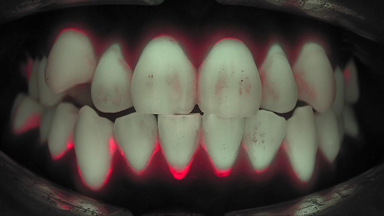
Dental plaque fluorescence raises an obvious question in the operatory: you see plaque every day, so why does only some of it glow red? Under white light, a patient's anterior teeth can look reasonably clean before a prophy. Capture the same arch under 405 nm blue light and a red band appears along the gingival margins and between the teeth. This post explains where that red comes from, why the eye and the bitewing cannot show it, and how to read it chairside. It is less about whether plaque is present and more about how long it has been there.
Why does plaque fluoresce red? The porphyrin story
The red comes from porphyrins made by bacteria. A porphyrin is a ring of four pyrroles. Hemoglobin in blood and chlorophyll in plants are the familiar examples. Some oral bacteria produce metal-free porphyrins such as protoporphyrin IX, and when these are excited by violet-blue light in the 400 to 420 nm range they emit strong fluorescence in the red part of the spectrum (Lee et al., J Biophotonics 2019).
One light, two signals: teeth glow green and bacteria glow red. Enamel and dentin absorb 405 nm light and re-emit green fluorescence. Porphyrin-bearing bacteria re-emit red. A single fluorescence image therefore carries two kinds of information at once: the condition of the tooth where green is lost, and the condition of the biofilm where red appears. The optics are covered in Quantitative Light-Induced Fluorescence: What 405 nm Shows.
The porphyrin producers are mostly late colonizers. Biofilm builds in layers. Early colonizers attach to the pellicle first; late colonizers arrive on top of them, and this later community is the one known to produce metabolites such as porphyrins. As a symbiotic biofilm drifts toward dysbiosis, the share of red-fluorescing bacteria grows.
Where does "three-day-old biofilm" come from?
In a lab model, red fluorescence showed up at about day three. Researchers grew microcosm biofilm from human saliva on bovine enamel discs and imaged it on days 1, 2, 3, 7 and 10. Red fluorescence intensity increased significantly through day 7 (p < 0.0001), and the point at which red became distinct sat around day three. That is the origin of the phrase "biofilm matured three days or more" (Kim et al., J Dent 2014).
Red tracked bacterial load, aciduric bacteria and enamel demineralization. In the same study, red fluorescence intensity correlated positively with total bacterial CFUs (r = 0.74), aciduric CFUs (r = 0.85), change in surface microhardness (r = 0.65) and lesion depth in the enamel (r = 0.82). The redder the biofilm, the more demineralized the enamel beneath it (Kim et al., J Dent 2014).
| Culture age | What fluorescence shows | State of the biofilm |
|---|---|---|
| Day 1–2 | Little or no red | Early colonizers dominate; not yet mature |
| Around day 3 | Red begins to appear | Late colonizers increase; porphyrin concentration rises |
| Day 7 and beyond | Red is distinct and widespread | Bacterial counts, aciduric bacteria and demineralization rise together |
Read the lab data as evidence of mechanism, not as a clinical clock. This was a microcosm biofilm on enamel discs, not a study of how many days plaque takes to turn red in a patient's mouth. What it establishes is the direction: red fluorescence reflects biofilm age and pathogenic potential, and later clinical studies point the same way.
Why can't the eye or the radiograph show it?
The eye sees plaque thickness and the radiograph sees mineral density. Neither shows biofilm maturity. A thin, colorless biofilm is hard to separate from the tooth surface under white light. A bitewing records differences in mineral density between tooth, bone and restoration, so a thin bacterial film does not register. Each exam answers a different question. None of them replaces another.
Disclosing shows where plaque is; fluorescence shows which plaque is old. Disclosing agents bind protein and stain new and old plaque alike. Two-tone disclosing colors thicker, older plaque blue. In a clinical study of 600 anterior facial surfaces in 50 adults, the area of red-fluorescing plaque (9.1%) was statistically similar to the blue-stained area (9.3%, p = 0.80) and different from the total red-stained area (31.6%). Red-fluorescing plaque also carried a higher proportion of *Prevotella intermedia* and *Streptococcus anginosus* (Han et al., Photodiagnosis Photodyn Ther 2016). In effect, fluorescence picks out the mature fraction without any dye.
| Method | What it shows | What it doesn't |
|---|---|---|
| Visual exam, white light | Thick plaque deposits, stain, calculus | Maturity of thin, translucent biofilm |
| Disclosing agent | Location and extent of plaque (rough age with two-tone) | Bacterial metabolism; patients dislike the stain |
| Radiograph | Changes in mineral density, bone, under restorations | Biofilm on the tooth surface |
| 405 nm fluorescence | Mature, porphyrin-bearing biofilm (red) and demineralized areas (green loss) | Lesion depth and extent; read with radiographs |
What bacteria live in red-fluorescing plaque?
Red plaque carried more periodontal pathogens and higher diversity. When paired samples of red-fluorescing and non-fluorescing plaque were collected from the same mouths and compared by 16S rRNA sequencing, red plaque showed greater microbial diversity and a significantly different community composition. Periodontopathic bacteria were significantly more abundant in red plaque, and fluorescence intensity related to the proportion of those bacteria and to the presence of gingival inflammation (Lee et al., J Biophotonics 2019).
Interdental red fluorescence lines up with gingival health. In 40 adults, interdental plaque between the first and second molars was collected with floss and imaged. Red fluorescence intensity and area were significantly higher in the gingivitis and periodontitis groups than in the healthy group (Guk et al., Photodiagnosis Photodyn Ther 2020). Red is not only a caries signal; it is a clue worth checking on the gingival side as well.
So red means "look here," not "disease here." A red area is an observation: mature biofilm is present. What it means, and what comes next, is decided by the dentist together with the visual exam, probing, radiographs and periodontal assessment. Like a traffic light, red says this is where we start care together.
A real case
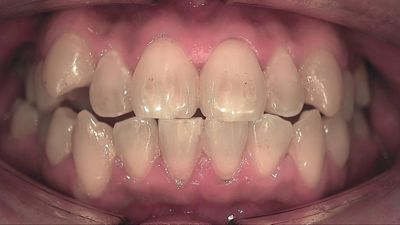
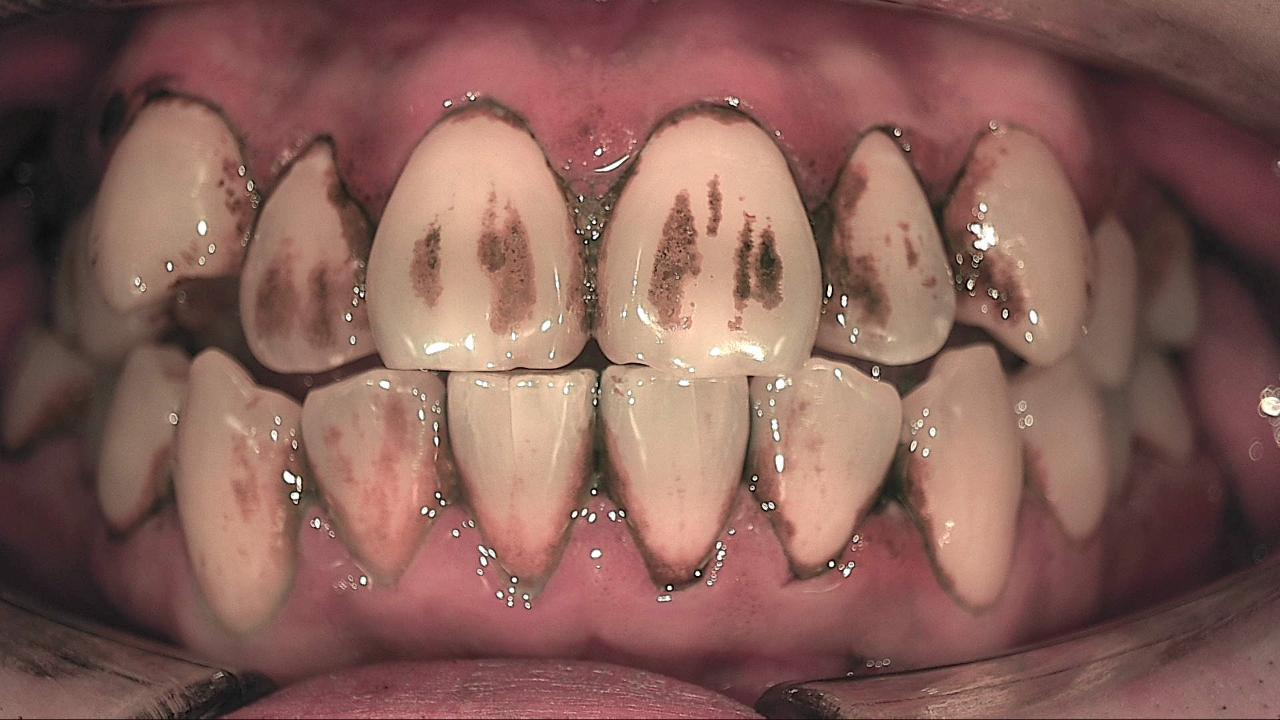
An arch that looks clean under white light reads differently with disclosing and fluorescence. This is a first visit before scaling. White light shows almost no plaque. Disclosing stains the cervical and interproximal areas broadly. Fluorescence narrows that down to a red band along the gingival margins, which is where mature biofilm has been sitting. That band, not the whole stained area, is the starting point for scaling and home-care coaching.
What's next
A red band is where Red Biofilm Care begins. Today's fluorescence image is recorded in LINKDENS and shared with the patient as a report. After scaling and home-care coaching, the same site is captured again under the same angle and light, and patient and clinician look at whether the red band has shrunk or stayed. For a practice owner, that turns "brush better" into "let's look at this red spot together at your next visit," a concrete reason to return. Repeated through QBLISScare, an invisible film of bacteria becomes the place where a patient's habits start to change.
FAQ
Q. If plaque fluoresces red, is it always caries?
A. No. Red fluorescence is an observation that mature, porphyrin-producing biofilm is present. It may be plaque sitting on the surface or bacteria inside a lesion, so clean first, look again, and read it with the visual exam and radiographs. The dentist makes the call.
Q. Does mature biofilm really take three days to turn red in the mouth?
A. Three days is when red fluorescence began to appear in a lab culture model. In a patient, saliva, diet and site all vary. The useful takeaway is the direction: the longer biofilm stays undisturbed, the redder it reads.
Q. Can fluorescence replace disclosing agents?
A. They answer different questions. Disclosing shows all plaque and its extent; fluorescence shows the mature fraction and is easy to repeat for visit-to-visit comparison without dye. Neither one replaces radiographs.
References
- Kim YS, Lee ES, Kwon HK, Kim BI. Monitoring the maturation process of a dental microcosm biofilm using the Quantitative Light-induced Fluorescence-Digital (QLF-D). J Dent. 2014;42(6):691-696. DOI
- Lee ES, de Josselin de Jong E, Kim BI. Detection of dental plaque and its potential pathogenicity using quantitative light-induced fluorescence. J Biophotonics. 2019;12(7):e201800414. DOI
- Han SY, Kim BR, Ko HY, et al. Assessing the use of Quantitative Light-induced Fluorescence-Digital as a clinical plaque assessment. Photodiagnosis Photodyn Ther. 2016;13:34-39. DOI
- Guk HJ, Lee ES, Jung UW, Kim BI. Red fluorescence of interdental plaque for screening of gingival health. Photodiagnosis Photodyn Ther. 2020;29:101636. DOI