
White Spot Lesion: No Red Yet? Recheck in a Month
A white spot lesion at the cervical third is a familiar judgment call in a recall exam. You can see it. You don't want to drag an explorer across it. The bitewing shows nothing yet. Do you restore today, or watch it? This post walks through what 405 nm Qray fluorescence adds at that moment, and where the chairside rule "no red yet — recheck in a month" comes from.
Why does a white spot lesion look white?
A white spot is the stage where demineralization runs just beneath the surface, before the surface breaks down. Incipient lesions keep the enamel surface continuous but look white because of a difference in refractive index. Advanced lesions lose that continuity and cavitate. Think of a sinkhole: we usually notice a sinkhole only after the ground collapses, yet the process was already underway underground.
The "underground" route is the microchannel network between enamel crystals. Enamel is not homogeneous like glass; it is an aggregate of microcrystals, and the channels between them carry organic acids, bacteria and minerals. That is why the surface layer can stay intact while mineral is lost right below it.
Caries progression can also be framed as a fire. Smoke comes first, then a small fire, then a large one. A large fire needs a fire truck; a sprinkler can handle a small one — and a smoke detector matters most.
| Fire stage | In the mouth | Response |
|---|---|---|
| Smoke | Mature biofilm | See it early, remove it, coach home care |
| Small fire | Incipient caries (white spot lesion) | Remineralize with fluoride and recheck |
| Large fire | Advanced caries | Restorative treatment |
What does a white spot lesion look like under Qray fluorescence?
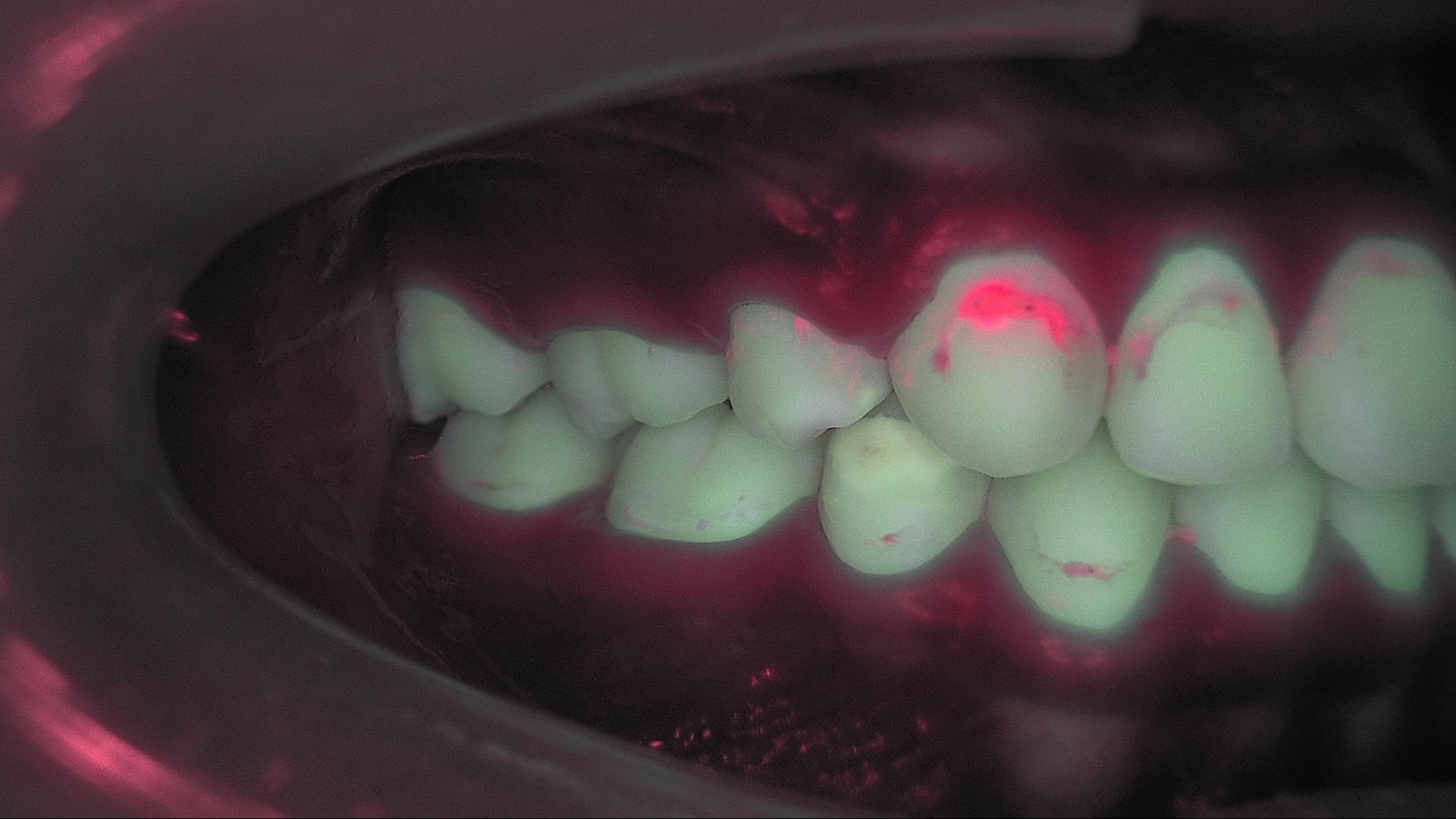
Dark with no red means pathogenic biofilm has no space to move into yet. Under Qray fluorescence, a surface lesion can scatter the natural fluorescence coming up from dentin, so the area may read darker — gray or brown. No red means calcium is leaving the surface and the structure is starting to change, but there is no gap for biofilm to colonize.
Red that survives cleaning means a physical space has opened and dysbiotic biofilm is moving in. If red remains after you polish the surface with pumice, acid from that biofilm will keep the lesion progressing.
How dark it reads is known to relate to how deep it goes. In an extracted-tooth study, fluorescence loss was measured alongside lesion depth by polarized light microscopy, and shallower lesions recovered more after fluoride treatment (Kim et al., Am J Dent 2013). That is one basis for estimating incipient lesion depth without sectioning the tooth.
| What you see under Qray fluorescence | What it means | Next step chairside |
|---|---|---|
| Darker area (gray/brown), no red | Subsurface demineralization; no gap for biofilm yet | Fluoride varnish plus high-fluoride toothpaste; recapture in about a month |
| Red that clears after cleaning | Mature biofilm sitting on the surface | Remove biofilm, coach home care, recheck |
| Red that persists after cleaning | Biofilm inside the lesion | Read with radiographs and the visual exam; decide on restoration |
Why wait a month before the red shows up?
When a surface lesion shows no red, a month of fluoride is worth trying first. One chairside approach is to apply fluoride assertively for about a month in these cases. In vitro, early lesions with fluorescence loss below a certain level often recovered by more than half after fluoride treatment (Kim et al., Am J Dent 2013). If a month of varnish and high-fluoride toothpaste does not bring the lesion back, that becomes the point to decide on a resin restoration.
If the red does not clear after cleaning and fluoride, you have a documented reason to restore. For lesions that sit right on the line between "treat" and "watch," this sequence gives you something more specific than either. You are not drilling today, and you are not putting it off indefinitely — you are scheduling a comparison of the same image in a month.
Lab data points the same way. An in vitro study (Kim, Photodiagnosis Photodyn Ther 2024) classified 44 non-cavitated lesions on extracted premolars and molars as active or inactive by red fluorescence intensity, applied 1.23% fluoride gel for 60 seconds, and stored them in artificial saliva for seven days. Recovery of fluorescence loss was 2.5 times higher in active lesions, a significant difference. This is an in vitro model, so read it as evidence of mechanism — active lesions respond to remineralization — not as a clinical outcome.
Developmental defect or early caries?
Not every white spot is caries, and the two call for different next steps. A white spot can be a developmental defect of enamel or an early carious lesion. A defect calls for monitoring and prevention; caries may need active intervention. Visual exams rely on color and opacity, explorers can damage weakened enamel, and radiographs have limited sensitivity for early enamel lesions.
Red fluorescence helps separate them. In a study of 704 teeth from 137 patients under age 20, green fluorescence loss grew and red fluorescence rose from sound teeth to developmental defects to caries (p < 0.001). White hypomineralization showed little red, while caries gave a clear red signal; the fluorescence difference between white hypomineralization and incipient caries was distinct enough to propose cutoff values for telling them apart (Lee et al., J Dent 2026).
Activity shows up in the red, not the green. In 34 primary molars classified as active or inactive by ICCMS criteria, green fluorescence loss did not clearly differ between groups, but red fluorescence was significantly higher in active lesions (p = 0.009) (Kim et al., Photodiagnosis Photodyn Ther 2022).
A real case
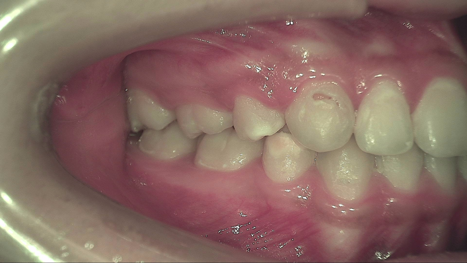
Under Qray fluorescence, the white areas you saw in white light split into those with a red signal and those without. The cervical white lesion on the upper tooth at center reads red in the Qray image, while the white areas on neighboring teeth show no clear red. That gives you two tracks: check whether the red persists after cleaning, and recapture the non-red areas under the same settings after fluoride.
What's next
The white-light and Qray fluorescence images you take today are recorded in LINKDENS and shared with the patient as a report. A month later, capture the same site under the same settings and put the two side by side: has the darker area brightened, and has any red appeared? For a practice owner, that is the moment "let's keep an eye on it" becomes "let's compare this picture together next month" — a clinical reason to come back. When that loop continues through QBLISScare, a small white spot becomes the starting point of lifelong care.
FAQ
Q. Does every white spot lesion need treatment?
A. No. A white spot can be a developmental defect or early caries, and early caries can remineralize with fluoride and home care. The dentist decides between monitoring, prevention and restoration, using Qray fluorescence images together with the visual exam and radiographs.
Q. If there is no red under Qray fluorescence, is the tooth fine?
A. No red only means biofilm has not found a gap yet — it does not mean there is no demineralization. Recapture darker areas under the same settings about a month after fluoride and compare.
Q. Does fluorescence replace bitewings for enamel demineralization?
A. No. QLF complements radiographs. Fluorescence shows early surface change and biofilm; radiographs show internal structure and extent. Read together, they give you a broader basis for the call.
References
- Kim HE, Kwon HK, Kim BI. Recovery percentage of remineralization according to severity of early caries. Am J Dent. 2013;26(3):132-136. PubMed
- Kim HE. Red fluorescence intensity as a criterion for assessing remineralization efficacy in early carious lesions. Photodiagnosis Photodyn Ther. 2024;45:103963. DOI
- Lee SJ, Choi HJ, Lee J, et al. Evaluation of developmental defects of enamel and differential diagnosis from dental caries using quantitative light-induced fluorescence technology: A cross-sectional investigation. J Dent. 2026;167:106546. DOI
- Kim CH, Bae K, Lee TY, et al. Assessment of dental caries lesion activity status using quantitative parameters obtained from the quantitative light-induced fluorescence method and difference of microbial distribution in primary molars. Photodiagnosis Photodyn Ther. 2022;39:102942. DOI