
Caries & Restorative
Early caries, proximal and occlusal lesions, white spots and secondary caries — seen through fluorescence imaging.
한국어 글 보기 →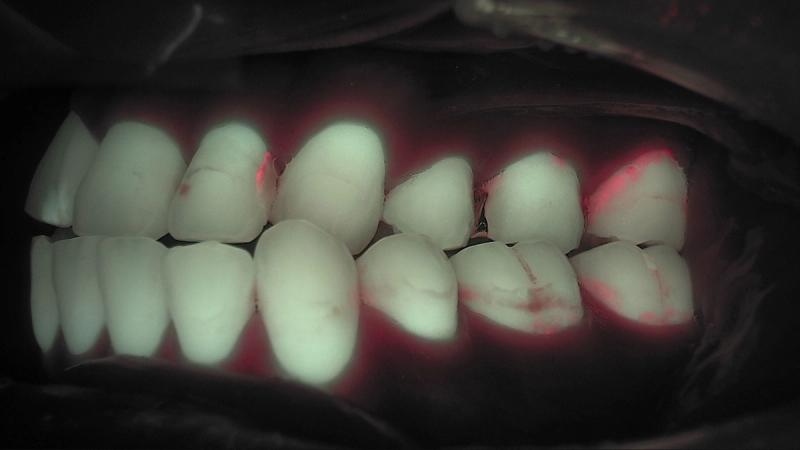
Early Caries Detection: Between the Eye and the Bitewing
Early caries detection is hard because the surface is still intact. Here is what the visual exam, radiographs and 405 nm fluorescence each show and miss, with meta-analysis figures, and the order to read them chairside.
Read →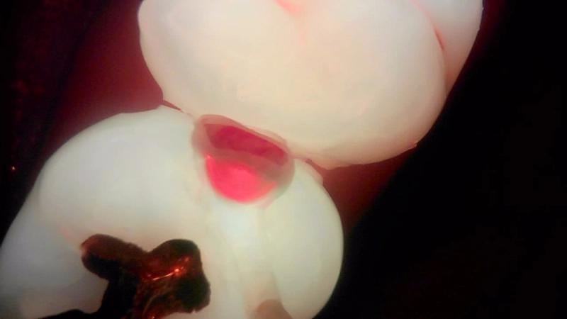
Interproximal Caries: Visual Exam, Fluorescence and Bitewing, Side by Side
Interproximal caries starts below the contact point, so the visual exam tends to see it late. We line up the visual exam, Qray fluorescence and the bitewing radiograph on the same surface and walk through what each view shows, what the evidence says, and a chairside workflow.
Read →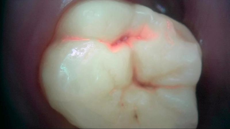
Stained Fissure vs Caries: Reading Occlusal Fluorescence
A dark fissure can be extrinsic stain or stain inside a lesion. Here is what the histology-validated research on stained pits and fissures shows, a 0–3 way to score occlusal fluorescence images, and how clinicians confirm "not caries" chairside.
Read →