
Stained Fissure vs Caries: Reading Occlusal Fluorescence
Stained fissure vs caries: it is one of the most common calls at a recall exam, and one of the least comfortable. A dark line in a molar's central fissure: watch it, or open it? And the patient often asks first: "Is that a cavity?"
This post walks through what to read on a fluorescence image of a stained occlusal surface, and how teams use the same image to explain why a particular dark spot may *not* need a drill. If you are new to the signal itself, start with Quantitative Light-Induced Fluorescence: What 405 nm Shows.
Why are stained pits and fissures so hard to read?
Color change in a fissure is not always a structural change. Chromogens from coffee, wine and tobacco stick to sound enamel; in a porous, demineralized lesion they soak in from the inside. Both look like a "brown fissure." Lee and colleagues (J Biomed Opt 2018) call the first noncariogenic discoloration and the second cariogenic discoloration.
Using stain as a criterion invites false positives. The same paper cites earlier work (Lussi, 1993) in which more than half of the teeth judged carious because of stained pits and fissures turned out to be sound.
On a fluorescence image, "dark" alone is not enough. Stain absorbs 405 nm light, so a stained fissure can look like it has lost fluorescence even with no demineralization underneath. In this study, histologically sound stained teeth still showed a median fluorescence loss of 62% compared with adjacent sound enamel. The authors concluded that fluorescence loss on its own is not reliable on stained occlusal surfaces, and proposed reading red fluorescence — which reflects bacterial metabolism — alongside it.
How does red fluorescence separate stain from caries?
Red fluorescence is not about color; it is about whether bacteria are living there. Porphyrins produced by bacterial metabolism get trapped in the porous structure of a carious lesion and glow red under 405 nm light. When the investigators imaged tooth cross sections, red fluorescence sat inside the lesion body, and its extent matched the demineralized area on polarized-light microscopy.
Against histology, red fluorescence tracked lesion depth most closely. The team imaged 62 extracted permanent teeth with questionable caries due to stained pits and fissures, then sectioned them. Twelve were sound (stain only) and 50 were carious — 43 in enamel, 7 into dentin.
| Parameter | Correlation with histology | Sensitivity / specificity | AUROC |
|---|---|---|---|
| Max red fluorescence gain | ρ = 0.80 | 0.96 / 0.83 | 0.94 |
| Max fluorescence loss | ρ = 0.76 | 0.80 / 0.92 | 0.91 |
The two numbers work as a pair. Red fluorescence was stronger at not missing caries (sensitivity); fluorescence loss was stronger at calling sound teeth sound (specificity). The authors are candid that this was an in vitro study, that values may be artificially high, and that clinical studies are still needed. Read it as evidence of mechanism, not as a chairside cutoff.
How do you score an occlusal fluorescence image?
A clinical scoring system gives your team a shared 0–3 language. Jung and colleagues (Photodiagnosis Photodyn Ther 2018) examined 791 teeth in 94 adults with ICDAS visual-tactile exams and QLF imaging, and built QS-Occlusal, a four-stage score based on fluorescence loss and red fluorescence.
| Score | What you see on the fluorescence image | Depth confirmed on histology |
|---|---|---|
| 0 | No fluorescence loss, no red | Sound |
| 1 | Faint loss or red within the pit or fissure | Outer half of enamel |
| 2 | Loss and red spreading beyond the fissure | Inner half of enamel |
| 3 | Red glow with a dark shadow | Into dentin |
Different readers landed on the same score. Intra-examiner agreement was ICC 0.94 and inter-examiner agreement 0.86, with AUROC from 0.807 to 0.976 across ICDAS thresholds. More than 43% of teeth scored ICDAS 0 on the visual exam had a fluorescence score of 1 or 2 — fluorescence change can show up before the surface looks different. Treat the score as a way to record what you see alongside the visual exam and radiographs, in words the whole team uses the same way.
How do clinicians confirm "not caries" chairside?
Clean first, then recapture. Chairside, the usual answer to "stain or caries?" is a sequence, not a single image. Remove plaque from the fissures with pumice, then recapture with Qraypen C — and a bitewing if needed. Red that was only on the surface comes off; when caries has spread beneath, the red can look wider after cleaning. For very early findings, factor in age and caries risk when deciding on timing.
If it is still there, look one step deeper. When fluorescence persists along a fissure after extra cleaning, one approach is a fissurotomy followed by a recapture; areas that turn more intense are more likely to be carious. The point is to avoid sealing over a hidden lesion.
"No red" is also a finding worth showing. A patient was sure an old filling had "turned black." The fluorescence image showed no red there: the dark area was amalgam showing through thin enamel, so no retreatment was needed. The conversation then moved to red areas that did need attention — and simply telling patients what is and is not caries can build trust. The same principle applies to a black-stained fissure in a child: check for red fluorescence, and if the lesion is not active, there may be no need to treat.
"No red" is not always the end of the story. When occlusal enamel is thick and a lesion has not reached dentin, an occlusal capture may show no red; adding a periapical radiograph can help. And some occlusal lesions show almost nothing on radiographs, which is why "visual exam → fluorescence capture → fluorescence analysis" is often kept as a fixed order. Fluorescence does not replace radiographs; it adds functional information between the visual exam and the film.
What does it look like in a real case?
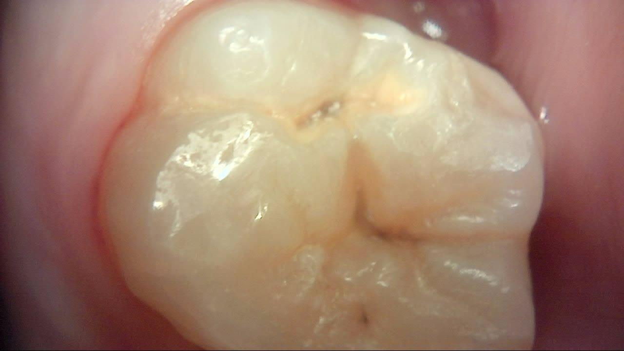
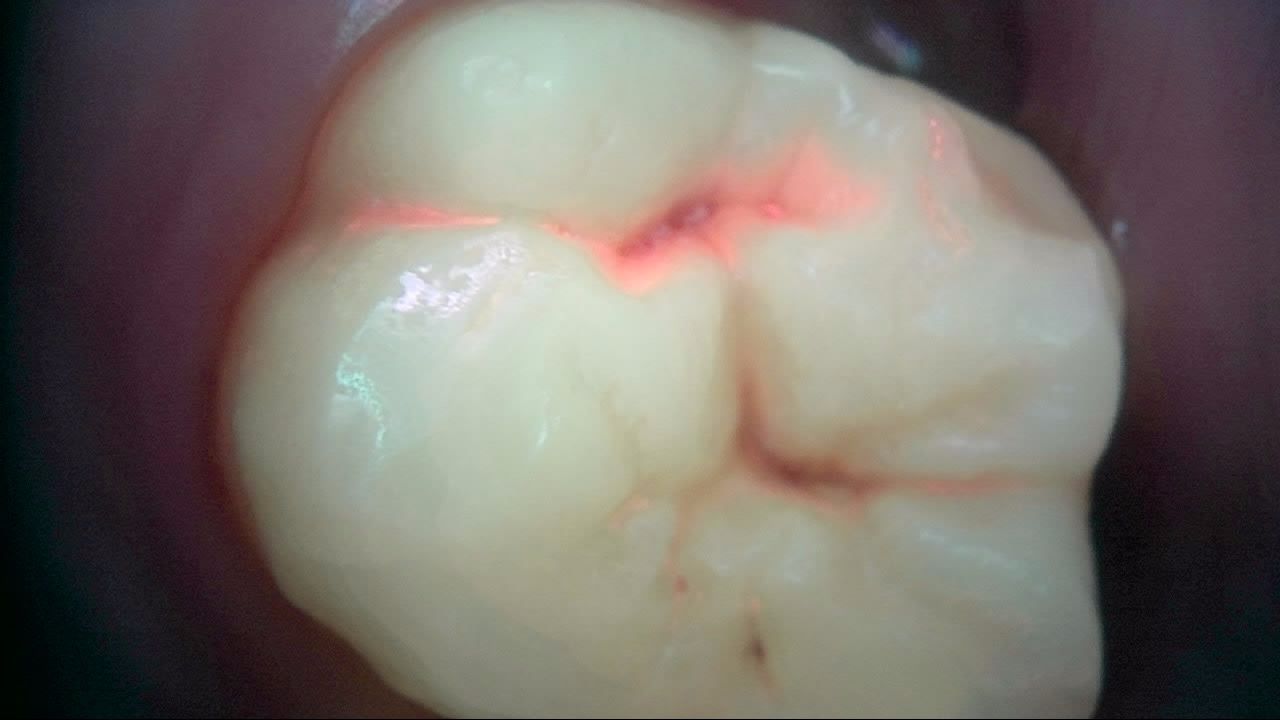
Under white light, this molar shows a small dark spot in the central fissure. Under fluorescence, red follows the fissure out from that spot — a starting point, not a verdict. Clean, recapture, and read it with the visual exam and radiographs.
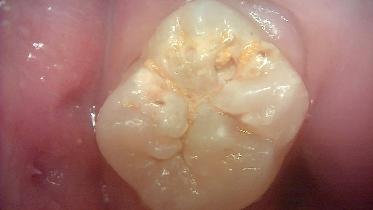
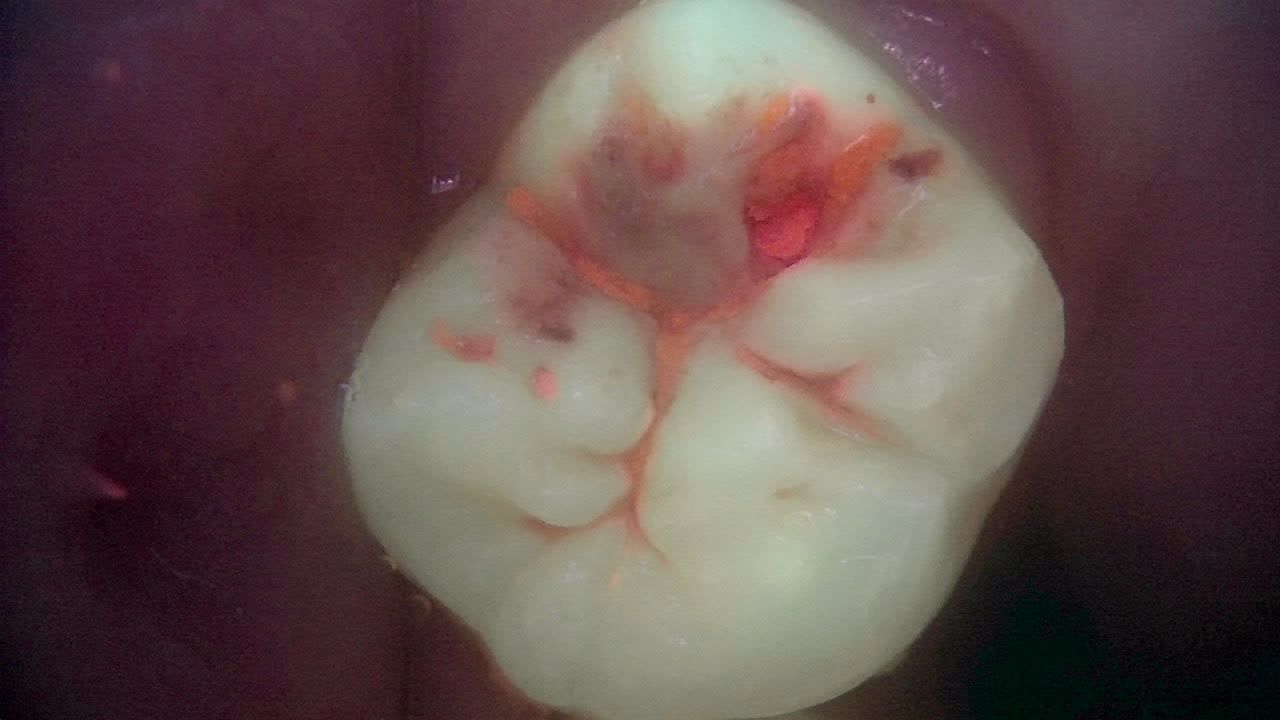
The second molar has broad yellow-brown discoloration around the fissures. Under fluorescence, red signal and a darker zone of fluorescence loss appear together — the same "discoloration" reads as two separate pieces of information. Whether and how far to treat stays with the dentist.
What's next
An occlusal fluorescence image should not end at the chair. Record it in LINKDENS and send it to the patient as a report, and a fissure you decided to watch becomes a fissure you can actually compare: same site, same settings, next visit — has the red grown or not? For the practice owner, that is a concrete clinical reason for the next appointment. Carried forward through QBLISScare, a single stained fissure becomes part of ongoing care.
FAQ
Q. Is a black spot in a pit or fissure always caries?
A. No. It can be extrinsic stain on sound enamel or stain inside a demineralized lesion. Check for red fluorescence, see whether it persists after cleaning, and decide together with the visual exam and radiographs.
Q. If a stained fissure looks dark on the fluorescence image, is that occlusal caries?
A. Not necessarily. Stain absorbs light and can mimic fluorescence loss. On stained occlusal surfaces, read fluorescence loss and red fluorescence together.
Q. Does red fluorescence mean I should restore right away?
A. Treat red as a signal to start managing together. Surface plaque comes off with cleaning, so clean and recapture first. Early findings may be monitored or managed preventively depending on caries risk; the treatment decision stays with the dentist.
References
- Lee HS, Kim SK, Park SW, et al. Caries detection and quantification around stained pits and fissures in occlusal tooth surfaces with fluorescence. J Biomed Opt. 2018;23(9):091402. DOI
- Lussi A. Comparison of different methods for the diagnosis of fissure caries without cavitation. Caries Res. 1993;27(5):409-416. DOI
- Jung EH, Lee ES, Jung HI, et al. Development of a fluorescence-image scoring system for assessing noncavitated occlusal caries. Photodiagnosis Photodyn Ther. 2018;21:36-42. DOI