
Early Caries Detection: Between the Eye and the Bitewing
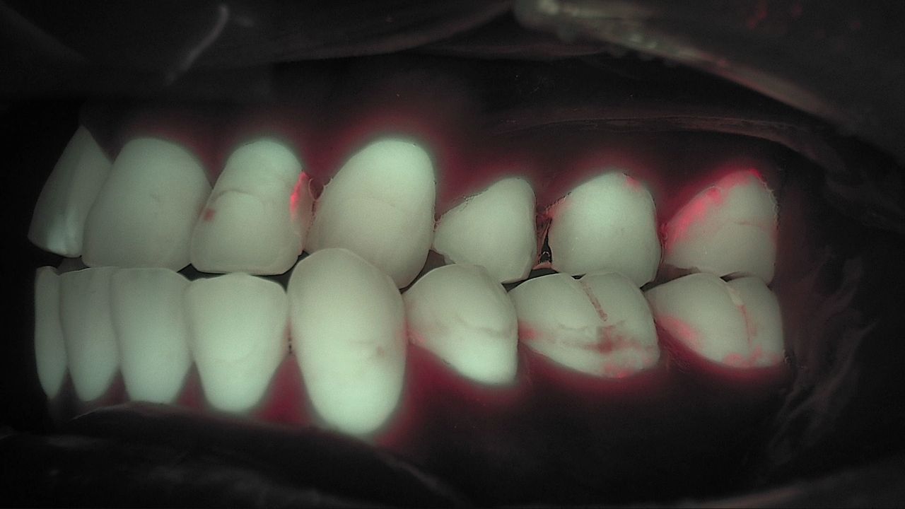
Early caries detection is where most of the hard calls in a recall exam live. A fissure looks a little dark, the explorer does not catch, and the bitewing is clean. Treating feels premature; watching feels vague, because you are not sure what you will compare against next time. This post walks through the three ways to confirm an early lesion, the visual-tactile exam, radiographs and 405 nm fluorescence, and what each one shows and misses, with the numbers from the literature. The short version: none of them is the answer on its own, and the picture gets clearer when you look at all three.
Why early caries sits between the eye and the bitewing
An early lesion is subsurface mineral loss under a surface that has not broken down yet. Enamel is an aggregate of microcrystals, and the channels between them carry acid out and mineral in. So the surface can look continuous while the layer beneath it is already demineralized. At this stage the lesion reads white or brown with no cavitation, and this is exactly where the treat-or-remineralize decision is made. The white spot stage is covered in White Spot Lesion: No Red Yet? Recheck in a Month.
Caries progresses when the balance between demineralization and remineralization tips. Acid from biofilm pulls mineral out; saliva and fluoride put it back, many times a day. If the balance stays tipped toward loss, the lesion deepens. If it swings back, the lesion stalls. Confirming early caries is less about "is there a hole" and more about "which way is this tooth leaning right now."
Method 1: Visual-tactile exam. First, but surface only
The visual exam comes first and is used most, but it reads the surface. It relies on color, opacity and texture, so lesions often become obvious only once they reach dentin. For non-cavitated lesions, reported sensitivity ranges from 0.2 to 0.96 and specificity from 0.50 to 1.00 depending on the examiner and the criteria (Oh et al., Sci Rep 2022). Explorers can damage weakened enamel, so they are used cautiously on early lesions.
Criteria such as ICDAS standardize the exam, yet two clinicians can still disagree on the same tooth. Case reports have noted that diagnoses based on traditional methods can differ under the same tooth condition, which is the argument for adding information rather than replacing the exam (Lee et al., Photodiagnosis Photodyn Ther 2019).
Method 2: Radiographs. Depth and extent, but blind to early lesions
Radiographs record changes in mineral density, which makes them strong for depth and extent. They are especially accurate for cavitated proximal lesions and dentin lesions. Bitewings remain the standard for proximal caries assessment, and nothing in this post suggests otherwise.
Sensitivity for initial enamel lesions, however, is low. A meta-analysis of 117 studies found that clinical radiographic detection of proximal lesions of any kind had a pooled sensitivity of 0.24 and specificity of 0.97, and for occlusal lesions a sensitivity of 0.35 and specificity of 0.78. The authors concluded that radiography is highly accurate for cavitated proximal lesions and that more sensitive methods could be considered for initial lesions (Schwendicke et al., J Dent 2015). A radiolucency appears only after substantial mineral loss, so "not visible yet" is not the same as "not there."
Method 3: 405 nm fluorescence. Demineralization and bacterial activity in one image
A fluorescence image shows where green has dropped and where red is glowing. Sound enamel re-emits 405 nm light as green fluorescence; demineralized enamel loses some of that green and reads darker. Mature biofilm and bacteria inside a lesion fluoresce red because of porphyrins. One capture carries both "where is the tooth demineralized" and "where are bacteria active." The optics are in Quantitative Light-Induced Fluorescence: What 405 nm Shows, and the red signal is explained in Dental Plaque Fluorescence: Why Mature Biofilm Glows Red.
It also gives an estimate of lesion depth. In extracted teeth, fluorescence loss in early lesions correlated strongly with lesion depth measured by polarized light microscopy (Kim et al., Am J Dent 2013). That is one basis for estimating the depth of an incipient lesion without sectioning the tooth.
Clinical studies report high accuracy on both proximal and occlusal surfaces. In a clinical study of 280 proximal surfaces assessed alongside the visual-tactile and radiographic exams, fluorescence parameters reached an AUROC of 0.86 to 0.90 at the dentin level, and a newly proposed fluorescence score scored 0.83 to 0.86 (Kim et al., Photodiagnosis Photodyn Ther 2017). In an occlusal study of 791 teeth in 94 subjects compared with ICDAS, a staged scoring system for non-cavitated lesions was developed from fluorescence images alone (Jung et al., Photodiagnosis Photodyn Ther 2018). In 878 primary tooth surfaces from 44 children, fluorescence parameters showed sensitivity of 0.72 to 0.91 and specificity of 0.74 to 0.96, similar to or slightly above the visual and radiographic exams (Cho et al., J Dent 2021).
A 17-study meta-analysis points the same way. The 2025 systematic review and meta-analysis reported pooled in vivo sensitivity and specificity of 0.86 and 0.82 for occlusal caries and 0.74 and 0.82 for proximal caries. The authors positioned fluorescence as an adjunct to the visual exam rather than a replacement, with particular value in monitoring lesion progression (Lee et al., Sci Rep 2025).
| Method | What it shows | Limits | Role for early caries |
|---|---|---|---|
| Visual-tactile exam | Color, opacity, surface texture | Subsurface is inferred; examiner variation | First gate. Picks the suspect sites |
| Radiographs | Mineral density, depth and extent | Low sensitivity for initial enamel lesions; biofilm invisible | Depth, treatment extent, cavitated lesions |
| 405 nm fluorescence | Demineralized areas (green loss) and bacterial activity (red) | Depth and extent need radiographs; surface plaque can overlap before cleaning | Early change, recapture under the same settings to compare |
What order chairside?
Pick the suspect site visually, read its condition with fluorescence, and confirm depth with a radiograph where needed. If fluorescence shows red, clean the surface and look again. Red that persists after cleaning raises the possibility of bacteria inside the lesion and is read together with the bitewing. Dark with no red means demineralization has started but there is no gap yet, so apply fluoride and recapture under the same settings to compare.
- Identify suspect sites with the visual-tactile exam.
- Capture the same sites under fluorescence and note green loss and red areas.
- If red is present, clean and recapture.
- Add a bitewing when depth or extent matters.
- The dentist decides between treatment, monitoring and prevention, and sets the next recheck.
Fluorescence does not mean the device makes the diagnosis. It adds one more image to the information the dentist weighs, and the decision is made together with the other exams. The meta-analysis authors drew the same line: an adjunct, not a replacement.
A real case
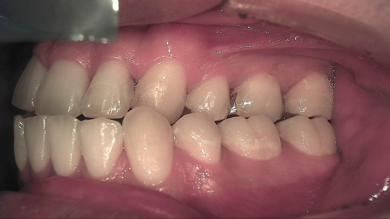
Surfaces that look smooth under white light split along their margins under fluorescence. Red fluorescence runs like a line along the cervical and interproximal areas, and a few teeth show darker patches near the occlusal surface. An image like this is not a conclusion that caries is present. It is the starting point: clean, see where red remains, and add a bitewing where it matters.
What's next
All three images, white light, fluorescence and the radiograph, are recorded together in LINKDENS and shared with the patient as a report. At the next visit the same site is captured under the same settings and placed beside the earlier record, so patient and clinician can see whether the darker area has brightened or red has appeared. For a practice owner, that turns "it's not bad enough to treat yet" into "let's compare this picture in three months," a clinical reason to come back. When that loop continues through QBLISScare, one early lesion becomes the start of lifelong care.
FAQ
Q. Do bitewings show every early caries lesion?
A. No. A radiolucency appears only after substantial mineral loss, so sensitivity for initial enamel lesions is low. For cavitated lesions and for judging depth and extent, radiographs remain the most reliable exam.
Q. Can fluorescence replace radiographs for caries detection?
A. No. Fluorescence shows early surface change and bacterial activity; radiographs show internal structure and extent. The meta-analysis positioned fluorescence as an adjunct to the visual and radiographic exams.
Q. Isn't the visual exam enough?
A. It is the first gate, but for non-cavitated lesions it varies widely with examiner and criteria. Reading fluorescence and radiographs alongside it broadens the basis for the call on the same tooth.
References
- Lee ES, Hwang J, Cheng L, et al. Diagnostic accuracy of quantitative light-induced fluorescence in detecting caries of various types and locations: a systematic review and meta-analysis. Sci Rep. 2025;15:39905. DOI
- Schwendicke F, Tzschoppe M, Paris S. Radiographic caries detection: a systematic review and meta-analysis. J Dent. 2015;43(8):924-933. DOI
- Oh SH, Choi JY, Kim SH. Evaluation of dental caries detection with quantitative light-induced fluorescence in comparison to different field of view devices. Sci Rep. 2022;12:6139. DOI
- Kim ES, Lee ES, Kang SM, et al. A new screening method to detect proximal dental caries using fluorescence imaging. Photodiagnosis Photodyn Ther. 2017;20:257-262. DOI
- Jung EH, Lee ES, Jung HI, et al. Development of a fluorescence-image scoring system for assessing noncavitated occlusal caries. Photodiagnosis Photodyn Ther. 2018;21:36-42. DOI
- Cho KH, Kang CM, Jung HI, et al. The diagnostic efficacy of quantitative light-induced fluorescence in detection of dental caries of primary teeth. J Dent. 2021;115:103845. DOI
- Kim HE, Kwon HK, Kim BI. Recovery percentage of remineralization according to severity of early caries. Am J Dent. 2013;26(3):132-136. PubMed
- Lee JW, Lee ES, Kim BI. Can red fluorescence be useful in diagnostic decision making of residual dentin caries? Photodiagnosis Photodyn Ther. 2019;26:43-44. DOI