
Intraoral Photo Comparison: Last Visit, Side by Side
Intraoral photo comparison is one of the simplest things a practice can add to a recall visit, and one of the most underused. Tell a patient "you're not brushing as well as last time" and they nod, but they have no way to check. Nobody remembers what their gingival margins looked like six months ago. Put last visit's Qray fluorescence image next to today's, and the conversation changes. This post covers what two images show that one cannot, what has to match for the comparison to mean anything, and the order that works chairside.
Why isn't one photo enough?
One image shows "now." Two show "change." If today's Qray fluorescence image shows red, mature biofilm is present. Whether that red has grown or shrunk since last time is something a single image cannot tell you. To decide whether the current approach is working, or something else is needed, you need a baseline. The red signal itself is explained in Dental Plaque Fluorescence: Why Mature Biofilm Glows Red.
The first step of proactive care is leaving a baseline. Proactive does not mean treating everyone sooner. It means looking at the current condition, recording a baseline, explaining what changes mean, and setting the next check. Today's image becomes next visit's reference, and that reference is what lets you separate, with evidence, the patients who need monitoring and coaching from those who need treatment.
It makes change visible that patients cannot feel. "I don't think you're flossing as well as you were six months ago" gives a patient nothing to verify. Six months ago next to today, and the patient spots where the red has grown before you point to it. The same works with parents of pediatric patients.
The evidence points the same way. The 2025 meta-analysis of 17 studies positioned fluorescence as an adjunct to the visual exam rather than a replacement, with particular value in monitoring lesion progression (Lee et al., Sci Rep 2025). In vitro, red fluorescence intensity has been used as a criterion for how early lesions respond to remineralization (Kim, Photodiagnosis Photodyn Ther 2024). The strength of fluorescence is less a one-time verdict and more the change over time.
What should you put side by side?
Pick one "let's look at this again" site per patient. You do not need to compare every tooth at every visit. Start with the places you have already promised to revisit: the cervical margins that read red today, a white spot you are watching after fluoride, a crack you have told the patient about, the margin of an old crown.
| Site to compare | What to look for across two images | Example recapture timing |
|---|---|---|
| Red fluorescence at cervical and interproximal areas | Has the red band shrunk, or stayed in the same place? | Next recall after prophy and home-care coaching |
| White spot lesion or early caries | Has the darker area brightened? Has new red appeared? | About one month after fluoride |
| A crack you have already disclosed | Has the line lengthened, or has red appeared along it? | Recall, or when symptoms appear |
| Margin of an older restoration | Has red appeared along the margin? | Recall; recapture after scaling |
The images can set the recall interval, too. Rather than one interval for everyone, some practices set three, four or six months based on how much biofilm is present. Patients with a lot of red come back sooner; when the side-by-side shows it shrinking, the interval stretches.
In one clinical case, a patient on a four-month biofilm-management recall had April and August images placed side by side. Much of the red seen in April was reduced by August after brushing coaching, and the patient saw the change for themselves.
Same conditions, or it isn't a comparison
A different date does not make it the same condition. A slight change in angle hides the cervical third. A different distance or focus changes how big the red area looks. Before putting two images next to each other, confirm they share the same site, the same angle and the same light. Keep the observation ("the red area is smaller") separate from the interpretation ("home care improved").
Ambient light matters more than most people expect. Operatory lights or window light mixing with Qray fluorescence can make the whole image read red. Put that image next to a clean one and the patient appears to have gotten worse when nothing changed. Dim the room lights and capture where ambient light is easy to block; that is where a valid comparison starts.
- Capture the same site from the same direction (fix an order, such as frontal, then left and right lateral).
- Keep the same distance, focus and light source.
- Cut ambient light from the operatory light and windows.
- Compare pre-cleaning images with pre-cleaning images, and post-cleaning with post-cleaning.
- Compare clinic images with clinic images. Use home images to show where to focus care, not as a like-for-like baseline.
What order works chairside?
Showing two images costs very little time. A fluorescence intraoral camera captures the white-light and fluorescence images with one press, so you are not doubling your photo count. A frontal view plus left and right laterals takes about a minute. Which device fits which job is covered in Qray Intraoral Camera Lineup: Which Device, When?.
The order is show, ask, decide together. Put last visit's image next to today's and ask before you explain. When the patient finds the difference themselves, the care conversation that follows gets much shorter.
- Capture today's images (white light and Qray fluorescence).
- Pull up the same site from the previous visit and place them side by side.
- Ask first: "What looks different between these two?"
- Name the observed difference and walk through home care again.
- Agree to look at the same spot next visit, and set when.
When the red has grown, don't blame. The image is not a report card; it is something you look at together. "This is a spot the brush isn't reaching, let's try it this way" moves the conversation to the next action, and makes next visit's comparison something the patient looks forward to rather than dreads.
A real case
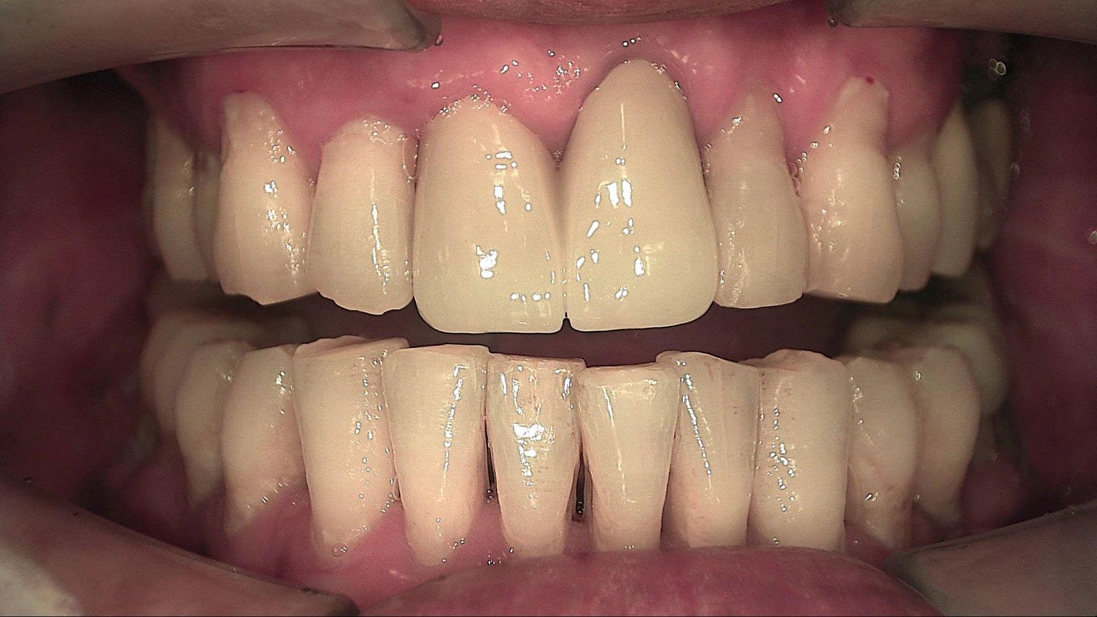
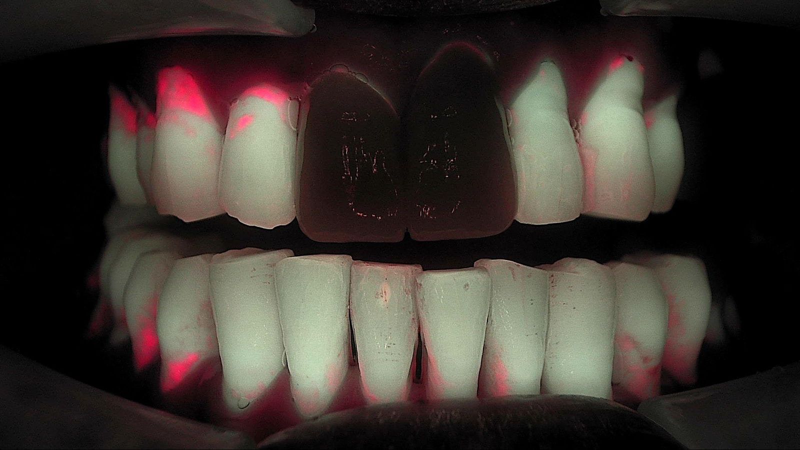
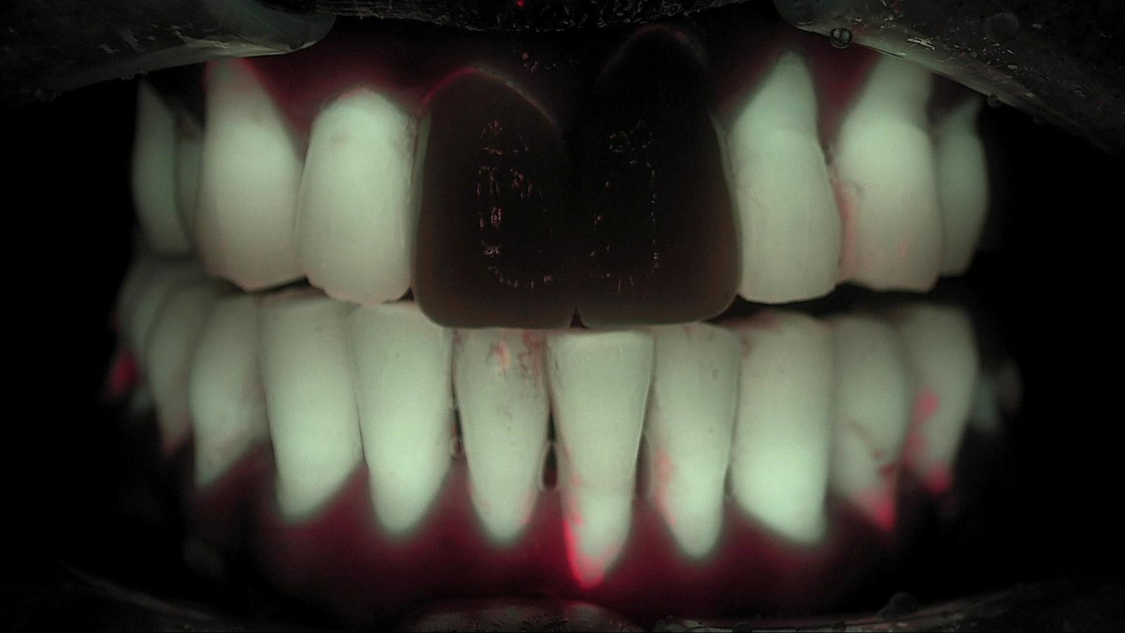
Same patient, ten months apart, both before scaling, both from the same angle. In the January 2024 Qray fluorescence image, red fluorescence is distinct along the cervical margins of the maxillary laterals and canines and between the mandibular incisors. In November, the red at those same sites is visibly reduced. The two maxillary central incisors are restorations, which fluoresce differently from natural teeth and read dark in both images. Side by side, the patient sees what changed before anyone explains it.
What's next
Today's images are recorded in LINKDENS and shared with the patient as a report, so the same site can be pulled up and placed side by side at the next visit. For a practice owner, that turns "we'll take another look next time" into "let's compare this picture in three months," a clinical reason to come back. When that comparison continues visit after visit through QBLISScare, a single photo becomes the record a patient uses to follow the changes in their own mouth.
FAQ
Q. How often should we do intraoral photo comparison?
A. There is no fixed interval; it depends on the patient. A patient with a lot of red fluorescence is compared at the next recall; a white spot under fluoride watch is recaptured in about a month. The dentist sets the next check.
Q. Can we compare a patient's home photos with clinic images?
A. Not on the same scale. Capture conditions differ, so home images are best used to show where to focus care. Comparisons of change should use clinic images taken under the same settings.
Q. Will showing comparison images increase return visits?
A. It cannot be guaranteed. What it does is make the reason for the next visit concrete, and there are practices that have found that easier to explain to patients.
References
- Lee ES, Hwang J, Cheng L, et al. Diagnostic accuracy of quantitative light-induced fluorescence in detecting caries of various types and locations: a systematic review and meta-analysis. Sci Rep. 2025;15:39905. DOI
- Kim HE. Red fluorescence intensity as a criterion for assessing remineralization efficacy in early carious lesions. Photodiagnosis Photodyn Ther. 2024;45:103963. DOI