
Root Caries in Older Adults: Spot It Early, Explain It
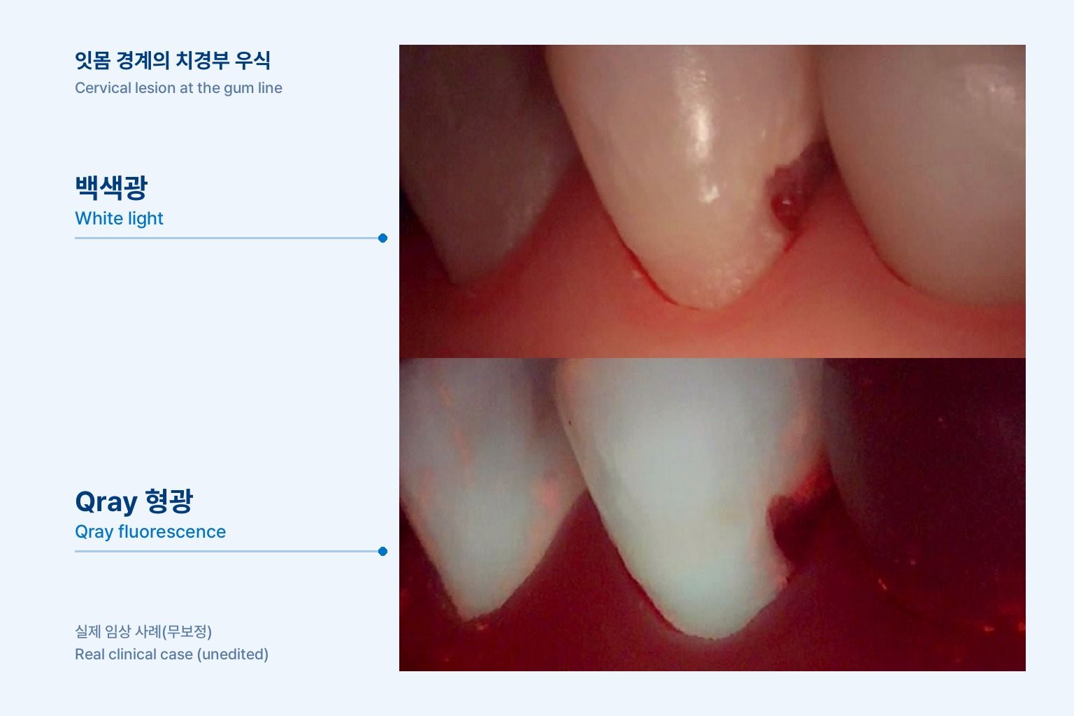
Root caries is the lesion that tends to slip through a busy recall schedule. Your hygienist finishes the prophy on a patient in her seventies, and on the lateral view there is a yellow-brown band at the cervical third of a premolar where the gingiva has receded. She mentions occasional sensitivity to cold. Is it stain, abrasion, the edge of an old Class V, or an active lesion? And how do you explain your decision in a way she will remember by the next visit? This article looks at root caries detection in older adults, what Qray fluorescence adds to the exam, and how to turn one image into a recare plan.
Why is root caries so easy to miss?
Exposed root surfaces are less resistant to acid than enamel, and they sit where plaque tends to stay. Once the gingiva recedes, cementum and dentin are exposed to the mouth. Plaque collects along the margin where the brush struggles to reach, and lesions often spread wide and shallow beneath it. Older patients tend to have more recession, and changes in dexterity or saliva can make home care harder.
The visual cues are ambiguous. Root lesions can look yellow or brown, and whether the surface feels hard or leathery is one of the main clues to activity. When abfraction, stain and the margin of an old composite overlap on the same surface, it is often hard to tell where the restoration ends and the caries begins. Read over plaque, the lesion can also look larger than it is.
How do the exam tools compare?
Visual-tactile, radiographic and fluorescence findings do not replace one another. Each answers a different question. The clinician decides activity and treatment with all of them together.
| Method | What it shows | Strength | Limit |
|---|---|---|---|
| Visual-tactile exam | Color, surface texture (hard or leathery), cavitation | Immediate, the basis for judging activity | Operator-dependent, and the finding is not saved the way an image is |
| Radiographs | Depth of proximal root lesions, bone level, relation to restorations | Shows depth and surrounding structures, and can be compared with later films | Buccal and lingual cervical lesions may be masked by overlapping tooth structure |
| Qray fluorescence image | Red fluorescence along the margin, areas darker than the surrounding root | No dye, and the image doubles as a patient explanation and a record | Must be separated from plaque and material fluorescence, and recent lesions may not glow red |
Red fluorescence marks sites where bacteria have settled, and darker areas relate to mineral loss. Red fluorescence takes time to develop after bacteria colonize, so the absence of red does not mean the surface is sound, and red does not automatically mean restore. Plaque itself glows red, so root surfaces should be re-imaged after the plaque is removed.
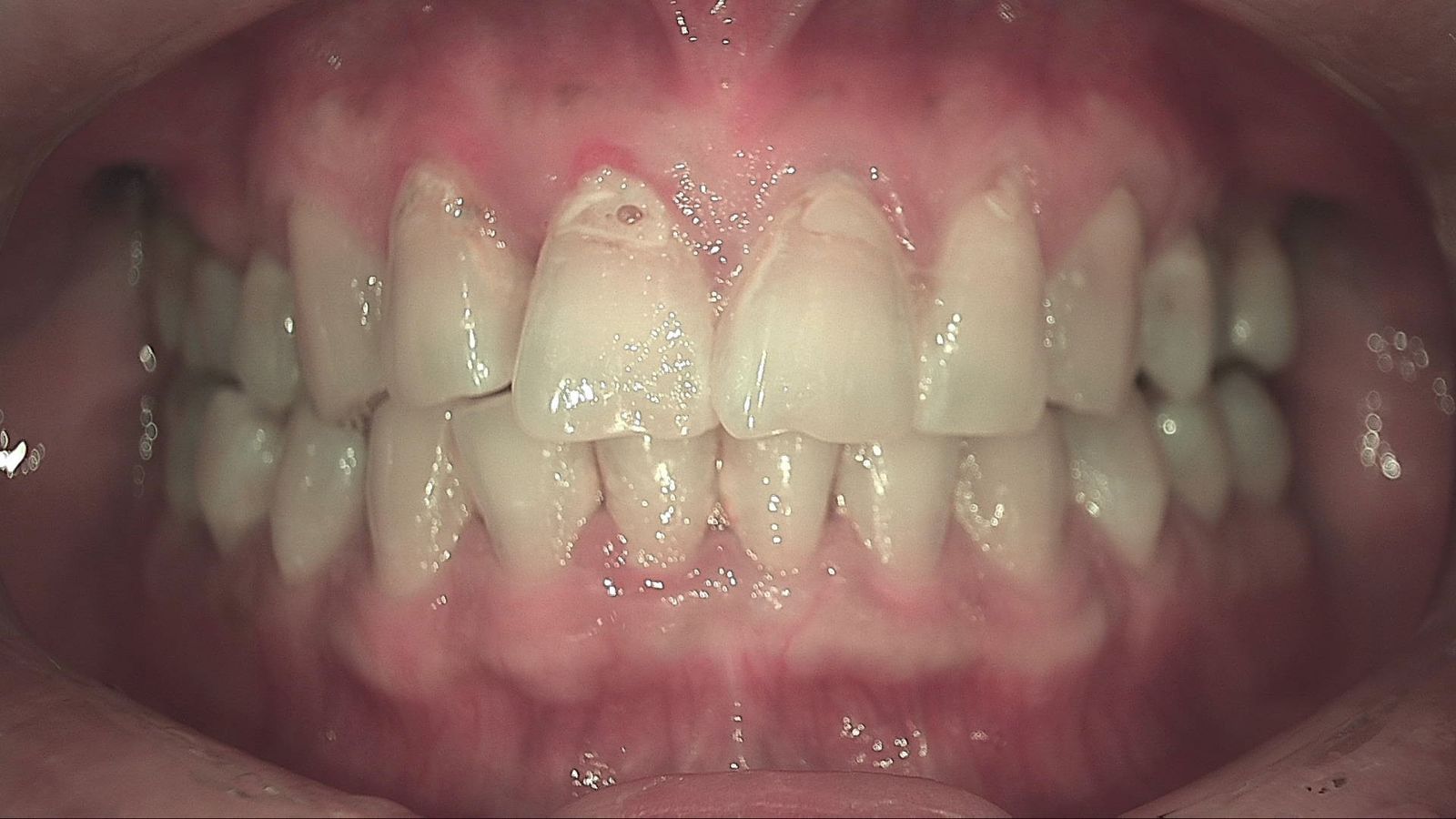
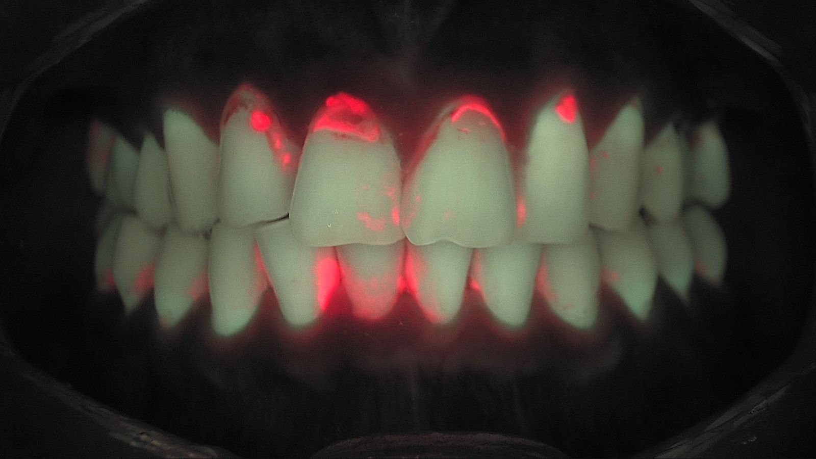
What does the research show about fluorescence on root surfaces?
One in vivo study imaged 46 exposed, non-cavitated root surfaces repeatedly with QLF. Three examiners imaged each surface three times, and both intra- and inter-examiner reproducibility were high. The amount of fluorescence loss correlated significantly with visual scores. The authors concluded that QLF can be recommended in addition to clinical evaluation for following the demineralization of exposed root surfaces over time.
A laboratory study found that fluorescence loss related to lesion depth. On 107 extracted teeth compared against micro-CT, leathery and cavitated root surfaces showed greater fluorescence loss than hard surfaces, and on non-cavitated surfaces fluorescence loss predicted lesion depth.
A randomized clinical trial in elderly participants used fluorescence to follow root caries management. Twenty-three participants with non-cavitated root lesions received different varnishes at regular intervals and were followed with fluorescence imaging for 12 months. No single varnish proved superior, but preventive care itself appeared to have a positive influence on lesion progress regardless of the varnish. Another study applied fluoride varnish weekly and was able to follow remineralization of intact root surface lesions at each visit.
These studies used research QLF systems and analysis methods. In your operatory, a Qray fluorescence image applies the same principle as a starting point for observing and comparing root surfaces, interpreted alongside your other findings.
A chairside sequence for recare
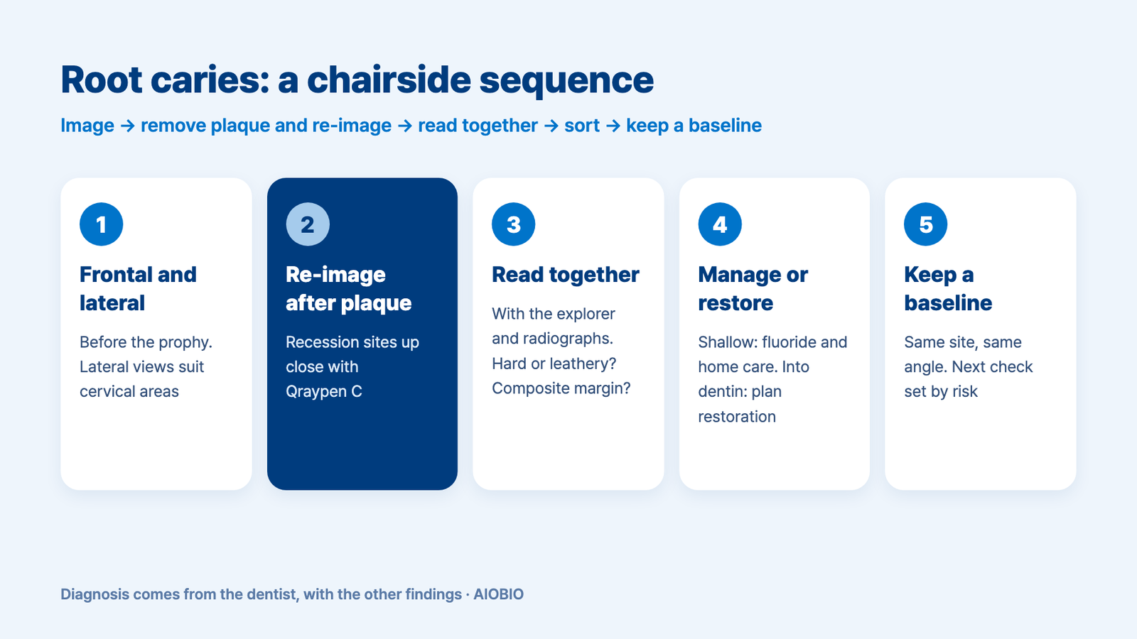
- Before the prophy, take frontal and left and right lateral images. Lateral views are well suited to abfraction and cervical caries.
- After plaque and calculus are removed, re-image recession sites up close with Qraypen C. For crowned teeth, angle up from below the cervical area toward the crown to see the margin.
- Read red and dark areas together with the explorer and radiographs. Check whether the surface is hard or leathery, and whether you are looking at a composite margin.
- Separate sites to manage from sites to restore. Shallow lesions can be followed with fluoride and home care, while lesions well into dentin are planned for restoration.
- Save the image taken at the same site and angle as the baseline, and set the next check by risk.
Show the patient where, why, and what you will look at next time. "This is where the gum has pulled back and the root is exposed. Even after cleaning, this edge still shows red, so we are going to work on it. Next visit we will take the same picture from the same angle and compare." In one clinical case, a patient who came in with a fractured premolar was shown the red area on the root surface and accepted the treatment plan after seeing it. Not every patient responds the same way, but an image anchors the explanation instead of replacing it.
What's next
Root caries care is a series of visits, not a single appointment. Today's images of the recession sites are stored in LINKDENS, delivered to the patient as a report, and can be placed side by side with images taken under the same conditions at the next recall. For a site you chose to manage with fluoride, the pair shows whether things are changing. For a site you restored, it becomes the reference for re-checking the margin and the surrounding root. It does not guarantee a return visit. It does give your team one consistent way to record and explain the gingival margin of every older patient, which is where continuous care through QBLISScare begins.
FAQ
Q. Is cervical caries the same as root caries?
A. They overlap but are not identical. Cervical caries is a broad term for lesions near the cementoenamel junction, while root caries refers to lesions in the exposed cementum and dentin after recession. Many lesions straddle both, so a lateral image helps you see the full extent.
Q. If a recession site glows red, should I restore it right away?
A. No. Clear the plaque and re-image first. If red remains, the clinician weighs surface texture, depth, radiographs and the patient's home care before choosing between monitoring with fluoride and restoring.
Q. How do I set the recall interval for root caries in older adults?
A. By risk rather than a fixed six months. Patients with many recession sites or recent lesions may need shorter intervals, with the same sites re-imaged from the same angle each time.
References
- Günther E, Park KJ, Meißner T, et al. Assessment of non-cavitated root caries lesions by quantitative light-induced fluorescence — An in vivo feasibility study. Photodiagnosis Photodyn Ther. 2020;30:101671. DOI
- Kreher D, Park KJ, Schmalz G, et al. Evaluation of quantitative light-induced fluorescence to assess lesion depth in cavitated and non-cavitated root caries lesions — An in vitro study. Photodiagnosis Photodyn Ther. 2022;37:102675. DOI
- Park KJ, Meißner T, Günther E, et al. Arrest of root caries with an adjuvant chlorhexidine-fluoride varnish over a 12-months observation period: a QLF-analyzed, placebo-controlled, randomized, clinical trial (RCT). Odontology. 2022;110(1):193-202. DOI
- Durmusoglu O, Tağtekin DA, Yanikoğlu F. Clinical evaluation of demineralization and remineralization of intact root surface lesions in the clinic by a quantitative light-induced fluorescence system. Lasers Med Sci. 2012;27(2):397-402. DOI