
Dental X-Ray Anxiety: Fluorescence and Radiographs, Together
Dental x-ray anxiety is a familiar moment in the operatory: "Can we skip the x-rays this time?" Sometimes the worry really is radiation. Just as often, the patient simply doesn't see why the image matters. A grayscale bitewing is your language, not theirs; to most patients it looks like a set of shadows. Making the invisible visible is, in the end, about helping patients understand their own condition so the next decision is one you make together.
This post looks at what Qray fluorescence and radiographs each show, where Qray fluorescence falls short, and a conversation order that keeps indicated radiographs on the table while respecting the patient's choice. The short version: the two exams give different information, and a site that needs a radiograph still gets one.
What's really behind dental x-ray anxiety?
Radiation concerns come from patients and parents alike. Plenty of patients feel uneasy about radiographic exams, and dental teams are careful about exposure, applying imaging criteria thoughtfully. With young children, sensor placement is difficult, and parents are often sensitive about radiation exposure.
Radiographs were never meant to be one-size-fits-all. One proximal caries study notes that, according to ADA guidelines, radiographic examinations should not be performed for screening purposes but on an individual basis, to minimize radiation exposure. The same paper notes that bitewings perform better than visual inspection for dentine-level proximal caries and serve as a criterion for operative decisions. That gives you an honest line for a worried patient: "We take radiographs where you need them, when you need them."
Hesitation is often about understanding, not radiation. "Is that really a cavity?" usually doesn't mean your call is wrong. It means the grayscale image doesn't speak to the patient. What patients push back on is often not the exam or the treatment itself, but what they can't make sense of. So a conversation with a patient who declines x-rays tends to go better when it starts with looking together rather than persuading.
Fluorescence and radiographs: what does each one show?
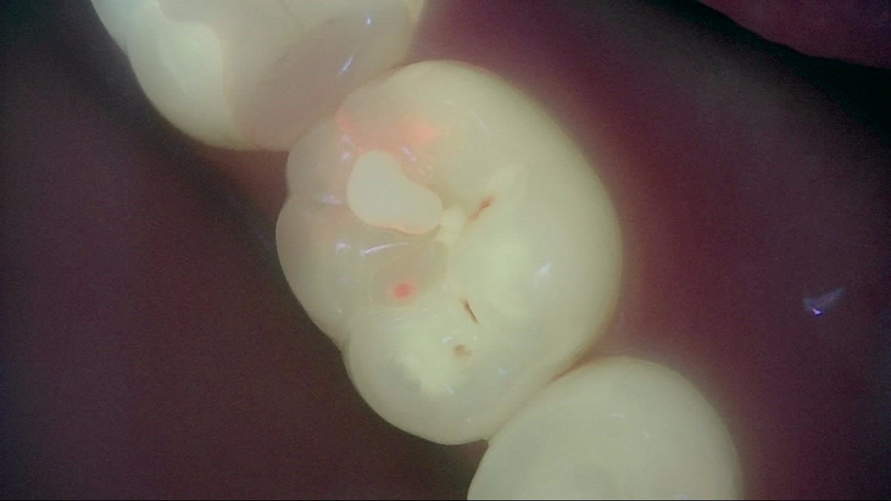
Qray fluorescence and radiographs show different information about the same tooth. Qray captures images with light from an LED source, not X-rays. Under Qray fluorescence, mature biofilm shows as red fluorescence, and demineralized surface areas look darker than their surroundings. Radiographs show how far a lesion extends into enamel and dentin, along with roots and bone. For why tissue glows red or goes dark in the first place, see Quantitative Light-Induced Fluorescence: What 405 nm Shows.
| What you're comparing | Dental radiograph (bitewing, periapical) | Qray fluorescence |
|---|---|---|
| Energy used | X-rays (ionizing radiation) | Light from an LED source; no X-rays |
| Mainly shows | Internal structure: lesion extent and depth, roots, bone | Surface information: red fluorescence from mature biofilm, darker demineralized areas, crack lines |
| Where it did better in research | Proximal caries | Occlusal caries, cracks |
| Hard to see | Biofilm distribution; early surface changes before they show as radiolucency | Early proximal lesions below the contact point; roots and bone |
| What the patient sees | Grayscale shadows that need your interpretation | Color on the tooth surface that patients can follow and ask about |
| Change over time | Recapture under the same conditions and compare | Recapture under the same conditions and compare |
In research, each method was stronger at different sites. In a retrospective study of 297 teeth from 153 patients, each imaged with both QLF and bitewing radiographs, fluorescence showed higher detection ability than conventional examination (visual inspection and bitewings) for occlusal caries and cracks, while bitewings detected proximal caries at a higher rate. The authors suggest that combining these complementary methods could reduce missed lesions, and call for a larger study to verify the protocol. These are results from a research setting; chairside, interpret them alongside your other exams.
Radiographs can be compared over time, too. Both exams can be recaptured under the same conditions and set next to the last visit. The difference is what you're comparing: with radiographs, changes in internal lesion extent; with Qray fluorescence, changes in the red and dark areas on the surface.
Where does Qray fluorescence fall short?
Early proximal lesions below the contact point may not be clear under Qray fluorescence. Most proximal caries start below the contact point, and when light comes in from the occlusal side, the sound marginal ridge weakens the fluorescence change that reaches the camera. The same proximal caries study noted that occlusal-direction fluorescence images are better suited to advanced lesions than early ones, and sensitivity and specificity were higher at the dentine level than at the enamel level. The retrospective study above likewise describes limitations for proximal caries because of the lesion's anatomical location.
A deep proximal lesion can look dark rather than red. Red fluorescence can be scattered or blocked by the overlying enamel. If a proximal area looks different from the surrounding sound enamel, even without red, treat it as suspicious and confirm with a radiograph.
Roots, bone and periapical areas are outside what Qray fluorescence covers. Those belong to periapical and bitewing radiographs, or other imaging as indicated. The same logic applies to panoramic images: rather than relying on a pan alone, confirm specific sites with periapicals or bitewings.
So a Qray fluorescence image with nothing on it doesn't mean a radiograph is unnecessary. Qray fluorescence adds information between the visual exam and radiographs. If symptoms or clinical findings call for a radiograph, take it. Which exam, and when, is the dentist's decision for the individual patient.
How do you talk with a patient who declines x-rays?
The Qray fluorescence screen can become the place where you and the patient see why a radiograph is recommended. In one clinical case, a Qraycam Pro image showed a subtle color difference at a proximal area. The clinician pointed to it and recommended a radiograph, and the patient agreed after seeing the area; looking back, the clinician felt the patient would likely have declined without the image. The radiograph then supported the call of proximal caries, and during treatment the lesion was removed a little at a time and re-imaged.
The flow can start from the radiograph, too. In another case, a patient who came in for a cleaning had a suspicious area on the panoramic image. After explaining it, the clinician captured a Qray fluorescence image, saw red fluorescence, added a periapical radiograph, judged the site to be proximal caries, and went ahead with treatment. The pan, the Qray fluorescence image and the periapical each added a different clue.
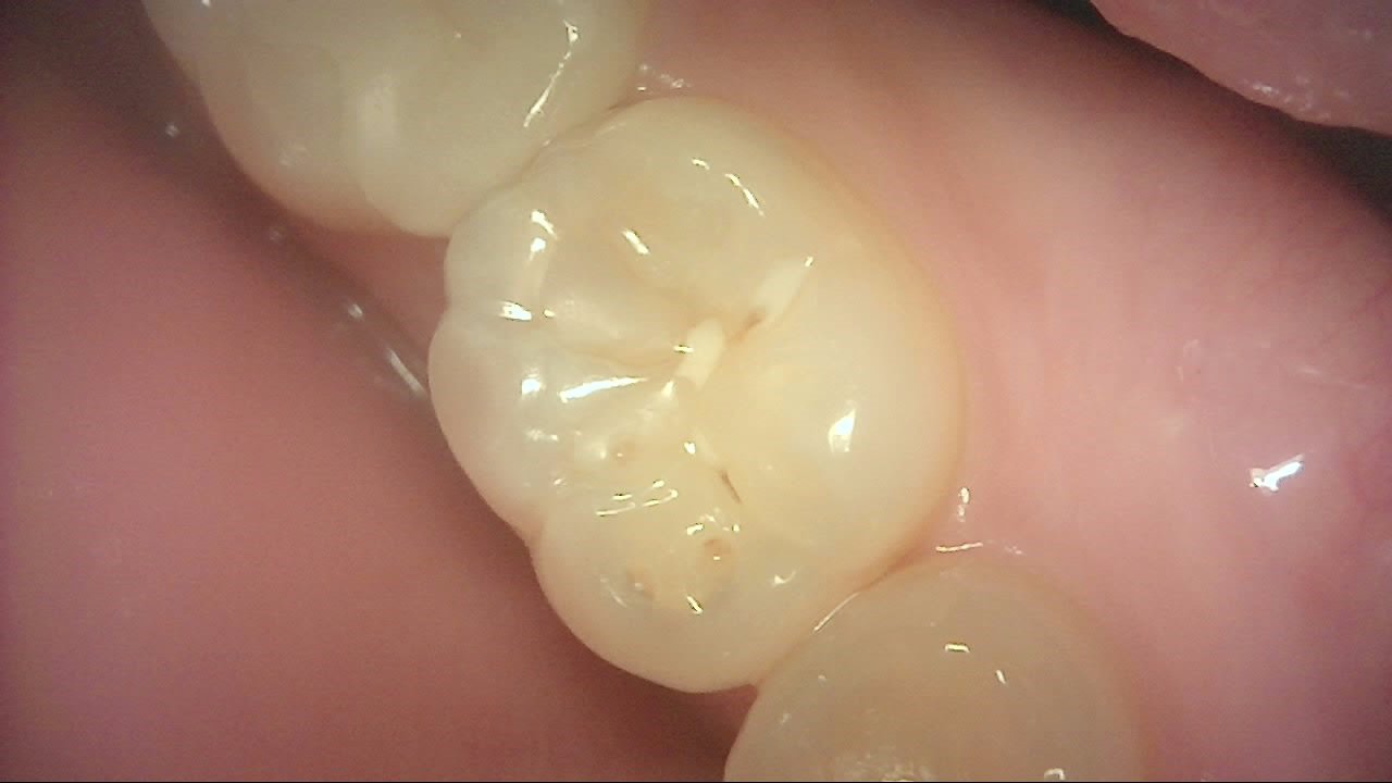
What shows on the surface and how far it goes inside are two different questions. These two images show the same tooth under white light and Qray fluorescence. The marginal ridge that looks smooth under white light shows a faint red signal under Qray fluorescence. The image tells you what's on the surface; it doesn't tell you how far anything extends into the proximal surface below. That's the kind of site you check with a radiograph as well.
A conversation order that works:
- Ask what the concern is first: radiation itself, cost or time, or not seeing the point.
- Look at today's tooth surfaces together under Qray fluorescence. Give the patient a moment to look and ask before you explain.
- Separate what the Qray fluorescence image shows from what it can't show.
- Explain which site needs a radiograph and why. Whether and when to take it is the dentist's call, based on the patient's condition.
- Record what was checked and what remains, and agree on when you'll look again.
For step 3, the wording can be this simple. "There's a red signal right here between these teeth. This image tells us what's on the surface, but not how far it goes inside. That's why I'd like to check this spot with a radiograph." Once the reason for the exam narrows down to one spot on the screen, it's easier for the patient to decide knowing what's being checked and why.
Some patients will still say no. Don't push. Explain what Qray fluorescence did and didn't show, and document that conversation. Make clear that nothing visible on the Qray fluorescence image doesn't mean everything inside is fine, and set a time to recheck so the conversation can continue at the next visit.
The same principle applies with parents. Starting with how well the child is brushing often makes the conversation easier. Early proximal lesions in primary molars can be missed under Qray fluorescence, though, so radiographs are still used when indicated. For more on sharing the screen with parents, see Caries in Children: Showing Parents What Primary Teeth Reveal.
What's next
Qray fluorescence images and the day's explanation go into LINKDENS and can reach the patient as a report. For a patient who postponed a radiograph, what was checked and what remains is on record, so at the next visit you can recapture under the same conditions, compare, and pick the conversation back up. For a practice owner, this turns time spent arguing over "x-rays or not" into time spent agreeing on what to check, and in what order. No one can guarantee consent or return visits, but you can help patients decide knowing what's being checked and why. As those checks continue visit after visit and carry into continuous care through QBLISScare, a single exam becomes the baseline for the next step in care.
FAQ
Q. Can I rely on Qray fluorescence alone for a patient who declines x-rays?
A. No. Qray fluorescence adds surface information. When you need to know what's happening inside a proximal surface, how deep a lesion goes, or what's happening at the root, confirm with radiographs. A Qray fluorescence image with no visible change doesn't mean a radiograph isn't needed. The dentist decides which exam, and when, for each patient.
Q. How do fluorescence and radiographs work as complementary imaging?
A. There's no fixed order. Sometimes a signal under Qray fluorescence leads to a bitewing to check the extent; sometimes a suspicious area on a pan is followed up with Qray fluorescence and a periapical. In one proximal caries study, the authors suggested fluorescence images can add information before radiographs are taken, while using bitewing scores as the reference standard.
Q. What should I tell an anxious patient about Qray?
A. Tell them plainly that Qray uses light, not X-rays. Light-based devices still have contraindications and precautions in their instructions for use: for example, patients taking photosensitizing medication or with a history of photobiological conditions such as erythropoietic protoporphyria or solar urticaria, and patients shouldn't look directly into the light. Check the labeling for your device and region. The accurate message isn't "this one test is enough," but "this helps you understand and choose the exams you need."
References
- Kim ES, Lee ES, Kang SM, et al. A new screening method to detect proximal dental caries using fluorescence imaging. Photodiagnosis Photodyn Ther. 2017;20:257–262. DOI
- Oh SH, Lee SR, Choi JY, et al. Detection of dental caries and cracks with quantitative light-induced fluorescence in comparison to radiographic and visual examination: a retrospective case study. Sensors (Basel). 2021;21(5):1741. DOI (Conflict of interest disclosed in the paper: one co-author is AIOBIO's CEO.)